Dear friends,
Well, we have arrived to case 25 after a bit more than a year. I hope that some of you have learned something and perhaps enjoyed the “game”. Let us continue…
Some days ago two smart radiology residents at Hospital Universitario Dr. Peset, Edgar Lorente and Alfonso Maldonado, presented me this case that has some interesting features.
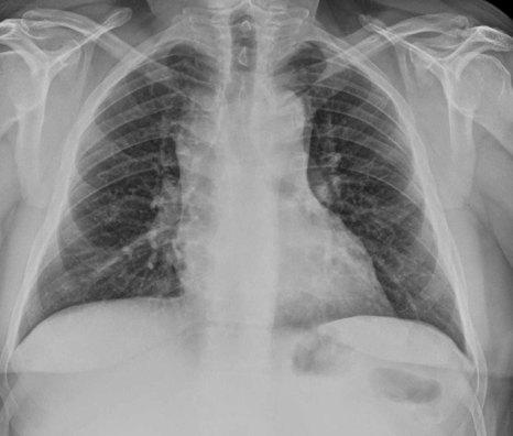
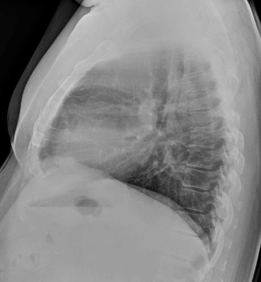
I will later show you the evolution, but let’s start with these images of this 34 year old man treated two years before of Hodgkin’s Lymphoma. The patient at the time of these images was asymptomatic, and the examination was part of the follow up.
What do you think. Is this recurrence of his Lymphoma?
Click here for the answer
Solution:
Well, not many people dared to give a diagnosis, but the ones who did were well oriented to something happening in the mediastinum. Indeed, what we see in the PA and lateral chest radiographs indicates that there is some mediastinal abnormality!
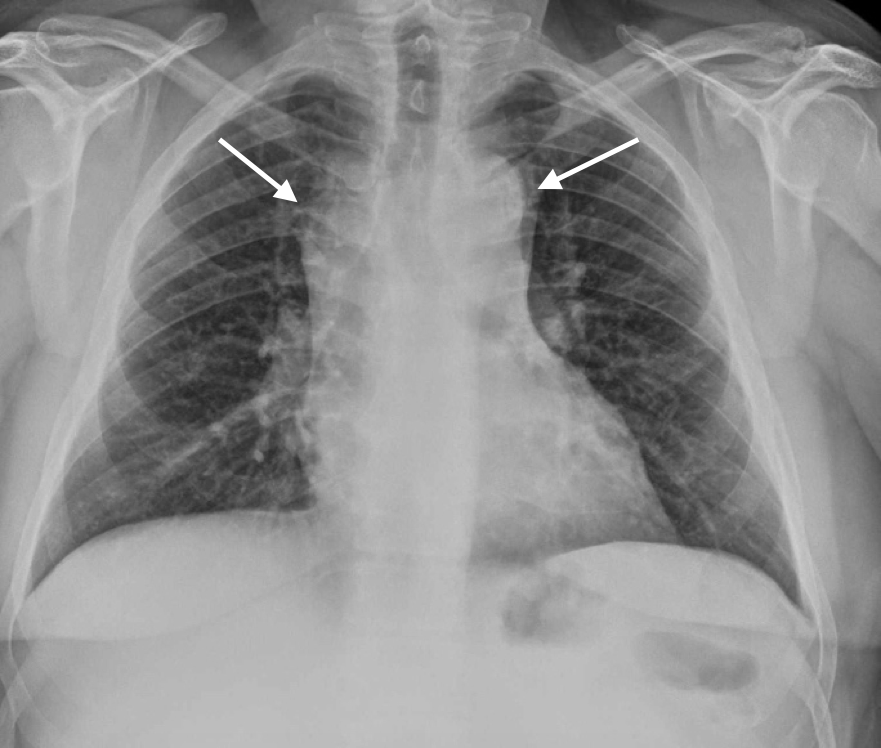
The PA radiograph shows a wide mediastinum (arrows) that seems to end in the clavicles indicating that the abnormality is anterior ( Cervico-thoracic sign).
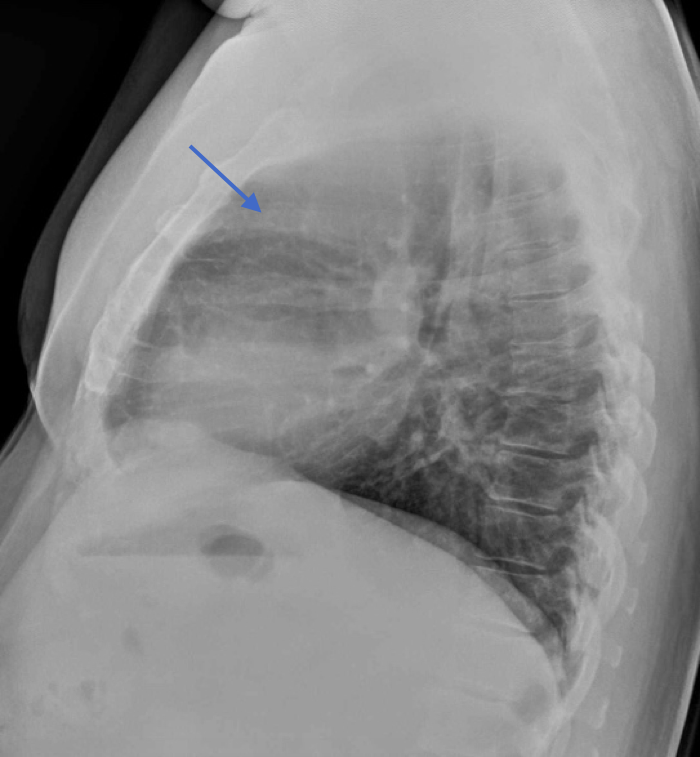
In the lateral radiograph, as in our case 24, you can see the there is occupation of the retrosternal space (Blue arrow) although the density seems a bit lower that what we could expect if there was a mass.
A CT was performed and showed that the “mass” was actually abundant fatty tissue.

The explanation in this case is that after treatment and a good response there has been a fatty replacement in the area where the lymphoma was present.
Fat replacement occurs in the chest often, either in the chest wall or in the mediastinum and may simulate other pathologies.
Some of you thought that this could be rebound thymic hyperplasia, and indeed this may occur in patients treated for lymphoma. In so called true thymic hyperplasia (rebound) the semiology is that of a mass that adopts the shape of an enlarged thymus. MRI in those cases with in and out of phase sequences will help with the diagnosis by showing a signal loss due to fat in the enlarged thymic gland.

Thymic hyperplasia in CT ( White arrow), “in phase” sequence showing high signal (yellow arrow) and “out of phase” sequence showing a loss of signal ( Red arrow.)
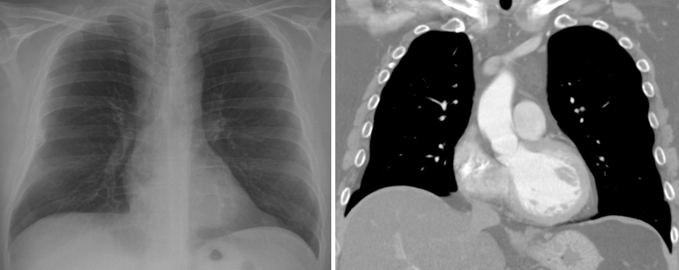
Mediastinal lipomatosis may occur also in obese patients such as in this other case. CT easily reveals the fatty nature of the mediastinal widening.
Teaching point: Fat in the mediastinum may appear and simulate pathology. CT or MRI can characterize it.


Rebound thymic hyperplasia after chemotherapy
Rebound thymic hyperplasia
enlargement of mediastinum perhaps complication of radiotherapy