Dear colleagues,
we are living dire moments. Our congress in Vienna has been postponed and many of us are subject to severe restrictions in our life, but this will not impede us to continue with our blog, and perhaps help you fill some time if you are isolated.

I would like to send you a case you already know, but with some new features stressed to me by Drs Maldonado and Lorente. Indeed I refer to our very last case (25) the man who had had Lymphoma and an accumulation of fat in the mediastinum: Remember?

Now, two years later the patient becomes symptomatic, with weight loss and fever.
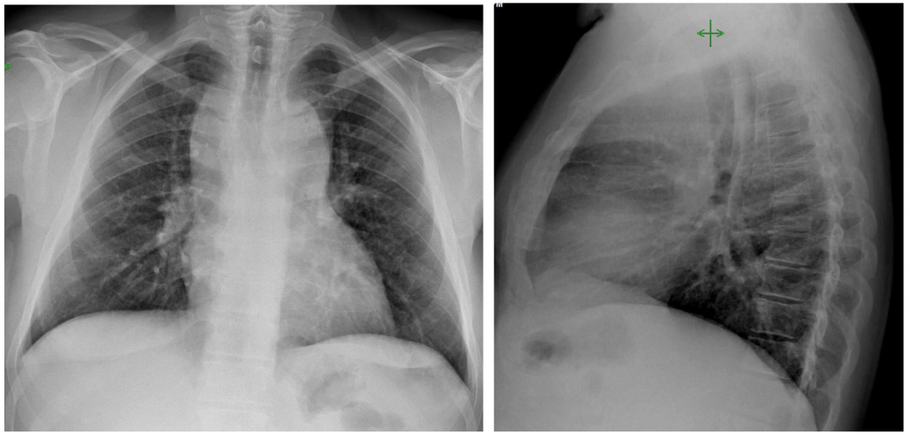
This is now his chest radiograph: What do you think?

Click here for the answer
Solution:
Well, this case was probably easier than others, but I thought that it could bring you a view of the different findings and scenarios that may happen along time in a patient.
Let’s recall:
1-Initial Images

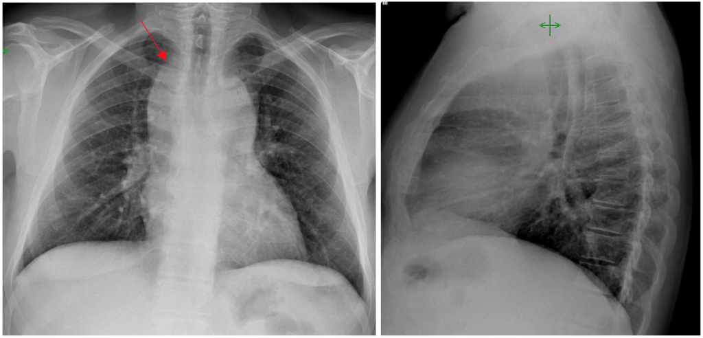
2- Two years later: The main finding is the widening of the anterior mediastinum with bulging in both sides and occupation of the retrosternal space. Notice that in the right side the limit is see surpassing the clavicle ( Red arrow) something that was not present in the previous study. This should raise the possibility of a mass growing there. Could it be more fat?

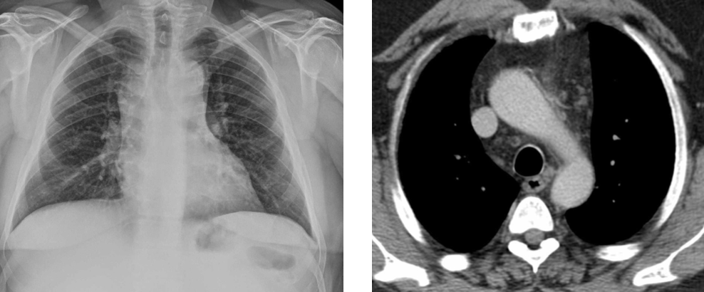
We obtained a CT:

The CT shows multiple adenopathies in the anterior, and middle mediastinum and some remaining fat in the most anterior part (White arrows). Also observe that the middle mediastinal adenopathies in the right have an interface with the lung ( red arrow) explaining the border seen in the PA radiograph.
Final diagnosis: Recurrence of Hodgking Lymphoma
Comment: Lymphoma is one of the most common causes of anterior mediastinal masses after thymic tumours. In contrast to Thymoma Lymphomas are usually bilateral masses, often “adapting” to the anatomic structures ( as enveloping) and extension to the middle mediastinum is common.


hi
my replay a thymome
I think it looks progressive increased opacity at superior mediastinum could be due to recurrent lymphoma, mediastinum lipomatosis, thymolipoma or thymic hyperplasia