Dear friends,
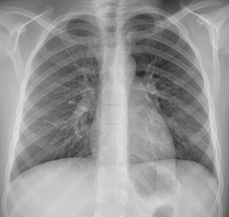
Warming up after the last case, here we are presented with a case of a 6-year-old boy with suspected pneumonia (cough and fever).
This case was shown to me by Dr. José Vizuete and Gregorio Martin; two former residents and now excellent radiologists at Dr. Peset Hospital.
What do you see from the image? Where is the pathology?
Just a little help in this case from my friend and colleague Dr José Vizuete
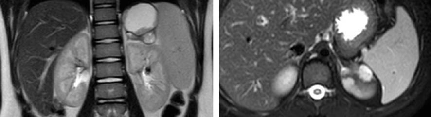
Two images of an abdominal MRI study in this young patient.
Any ideas?
The solution will come soon…
Click here for the answer
There are no signs of pulmonary infection, but if you look closely at the left sub phrenic region there is a curvilinear calcification (white arrows).
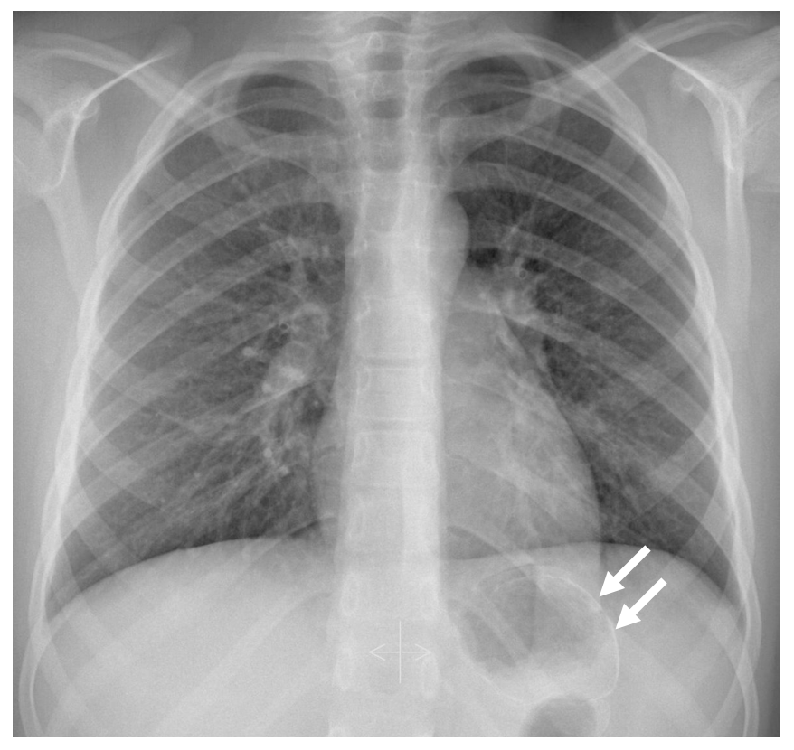
An abnormal calcification in the abdomen in a child should alert and move you to discard abdominal pathology.
MRI showed a left adrenal mass partially cystic but with nodules that enhanced with contrast.
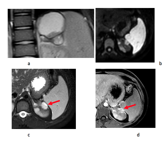
MRI: Coronal T2, axial STIR, axial diffusion and axial T1 with contrast. The lesion shows peripheral areas of hypointensity due to the calcified capsule (a), restricted diffusion (b) and peripheral hyperintense nodules (c) that enhance in the T1 sequence (d): (red arrows).
DIagnosis: Ganglioneuroblastoma.
Teaching point: Upper abdominal pathology can be seen in chest radiographs,
especially when the lesion contains calcium.
Comment: Hydatid cyst was initially included in the differential diagnosis, but the presence of enhancing nodules discarded this choice.
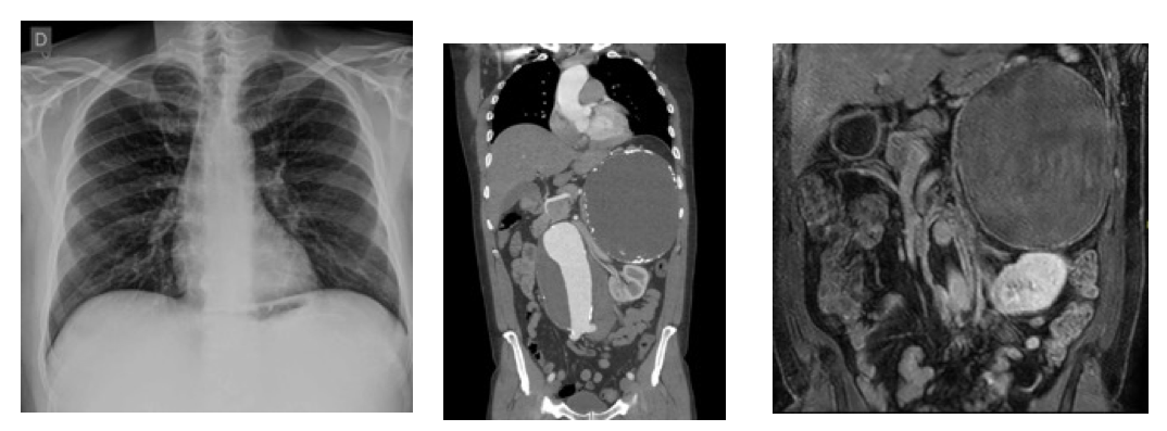
Suprarenal Hydatid cyst ( Left subphrenic curvilinear calcification). MR: Peripheral rim of enhancement but no enhancing nodules).


