José Vilar and Friends Case 31 (Update: Solution)

In this photo: Dr. Villanueva in the Caceres Corner
Hello friends.
I am back after a short medical leave.
Thanks to my friend Dr Alberto Villanueva (Sussex) I can show you an interesting case to test your skills once more.
17 yr. Fever and sore throat.
Let me know what you think in the comments!
Click here for the answer
This case was provided by my good friend Dr Alberto Villanueva-
The correct diagnosis was made by the majority of those who “dared” to send an answer although we know that many more people looked at the case. Anyway, let me congratulate Carmen Gassent, Andrii Veselyi, Mahdy, Elnaz Tabibian,Ahmed Kamalaldin, Lee, Selçuk Akkaya for making this diagnosis. Wow you are good! (But of course, I was kind enough to show the cervical CT which, you must admit that it was the clue…)
Let’s see the findings

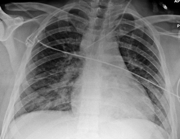
AP erect Chest radiograph: Some pulmonary opacities especially in the left lower lobe.

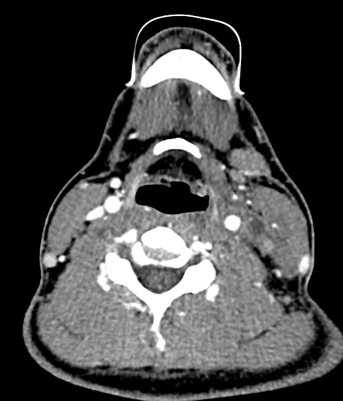
Cervical Contrast CT: Axial: There is an occlusion of the left internal Jugular vein (Yellow Arrow). No collections are seen.
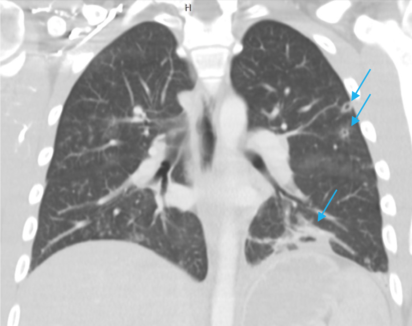
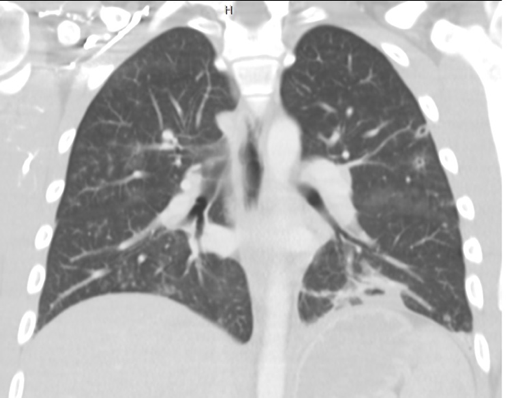
Thoracic CT. Coronal: Large left lower lobe cavitated consolidation and two small cavitated nodules in the left upper lobe. (Blue arrows)
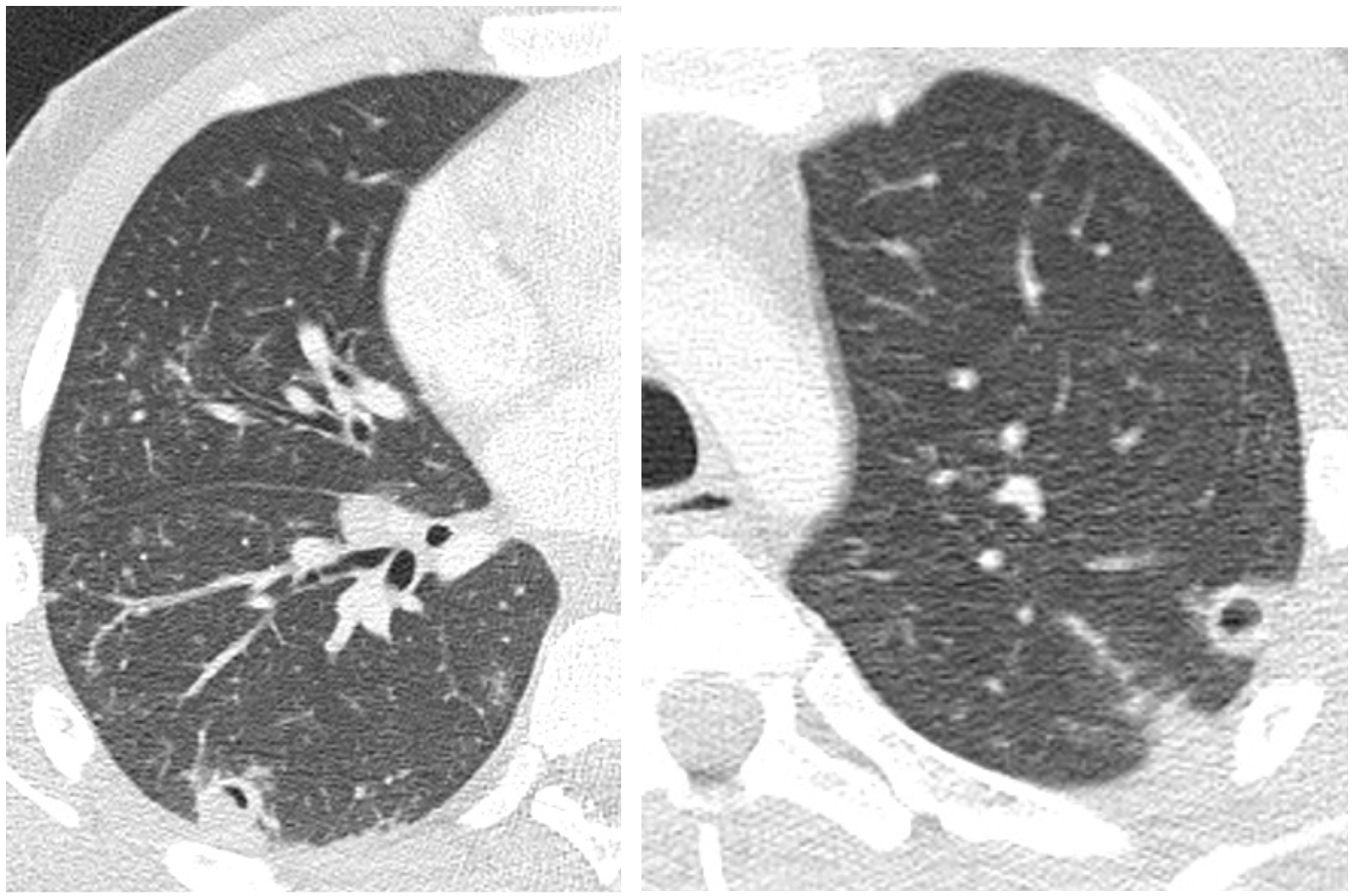
CT axial images not shown initially: Other cavitated nodules are seen in both lungs.
Diagnosis:
This case of a 17-year-old young man is clinically an acute process. The presence of multiple cavitated nodules in both lungs may suggest several conditions. In an acute clinical scenario, some diseases such as neoplasms or vasculitis are excluded.
Well, for those who did not know this pathology or forgot about it, here is a case of Lemierre’s disease: Oropharyngeal infection and subsequent thrombosis of the jugular vein with pulmonary septic emboli.
This disease was described originally by Andree Lemierre, a bacteriologist who in 1936 described it in 28 children , although in 1900 this type of infection had already been described by Cade and Courmont.
The main cause is in 80% of the cases Fusobacterium necrophorum although other bacteria may be associated.
This disease is less frequent since the advent of antibiotics, but may still appear and we should suspect it in the presence of oropharyngeal infection and pulmonary cavitated nodules in children or young adults.
Ultrasound and especially Doppler will identify the thrombus (hyperechogenic) in the internal jugular vein, other findings such as cervical abscess may be associated.
Contrast CT, covering the neck and the thorax is the most useful imaging procedure since it will show the thrombosed vein and the pulmonary cavitated nodules.
Lemierre A: On certain septicaemias due to anaerobic organisms. Lancet 1936, 1:701-703.
Meuwly JY, Lepori D, Theumann N et-al. Multimodality imaging evaluation of the pediatric neck: techniques and spectrum of findings. Radiographics. 2005. 25 (4): 931-48. https://doi.org/10.1148/rg.254045142


? Wegener’s granulomatosis
Wegener’s granulomatosis
Lemierre syndrome
Pequeñas imágenes nodulares, algunas cavitadas en ambos campos pulmonares (embolias sépticas), trombosis de la VY
Síndrome de Lemierre
thankyou for sharing:
1. chest xray:
– retrocardiac opacity
– few nodular densities in middle and right lower zone
2. ct chest :
– left lower lobe consolidation
– ground glass densities in middle and right lower lobe
– tiny suspicous ring like nodules in left upper lobe near periphery / subpleural region ? cavitating nodules
3. IV contrast neck ct ( above level of cords)
– left submandibular discrete oval lymph node with loss of fatty hilum
– tiny cystic area near left carotid vessel and below left sternocleidomastoid muscle ? necrotic lymph node ? cyst
differential diagnosis
? inflammatory process
? autoimmune disorder
can u tell me more about relation auto immune disease in these case please
Lemierre’s syndrome
Left internal jugular vein thrombosis secondary to left internal jugular catheter
Pulmonary septic emboli
Lemierre syndrome
Lemierre’s syndrome
Lemierre’s syndrome :
Cavitary pulmonary nodules due to septic emboli + Left side internal jugular vein thromophlebitis
IJV Thrombosis
Septic pulmonary Emboli
Lemierre’s syndrome
Thrombosis left IJV with cavitation lesions in left upper lung lobe
Lemierre syndrome – thrombophlebitis of the left internal jugular vein, pulmonary septic embolia
wow, most of you are well in the track.
Lemierre syndrome. Thrombophlebitis of the Internal al jugular vein, Septic pulmonary emboli
Cervical lympadentis (level II)
Peri adenitis around carotid space
Anterior jugular & internal jugular thrombophlebitis
Cavitaring nodules in periphery and consolidation left lower lobe
Constellation of findings Leimierre`s syndrome
Lemierre syndrome