José Vilar and Friends Case 33 (Update: Solution)

Hello Friends,
here I am trying to initiate a new course with fresh cases. I hope that all of you are well, and doing your best during this horrible pandemia.

My first case was shown to me a couple of days ago by Dr Teresa Lago Muñoz, a young radiology resident at Hospital Dr Peset.
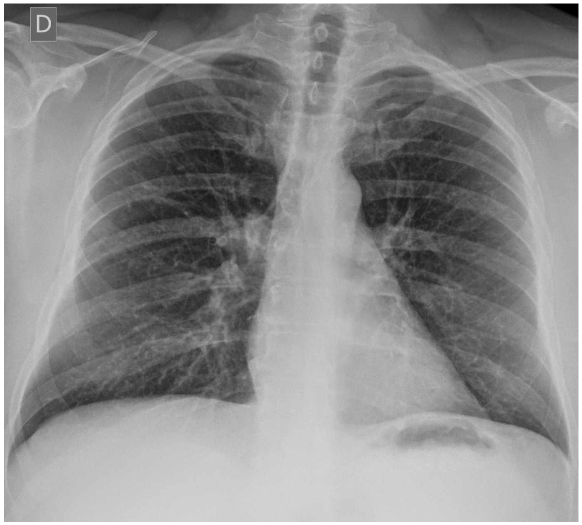
Dr Lago tells me that this is a 48 year-old man, heavy smoker for the last 30 years.
What do you think? What would you do? (No previous images were available).

Click here for the answer
This first case is very simple to warm up for future more complicated cases but yet this type of cases are important in our practice. Let´s see…
Solution.
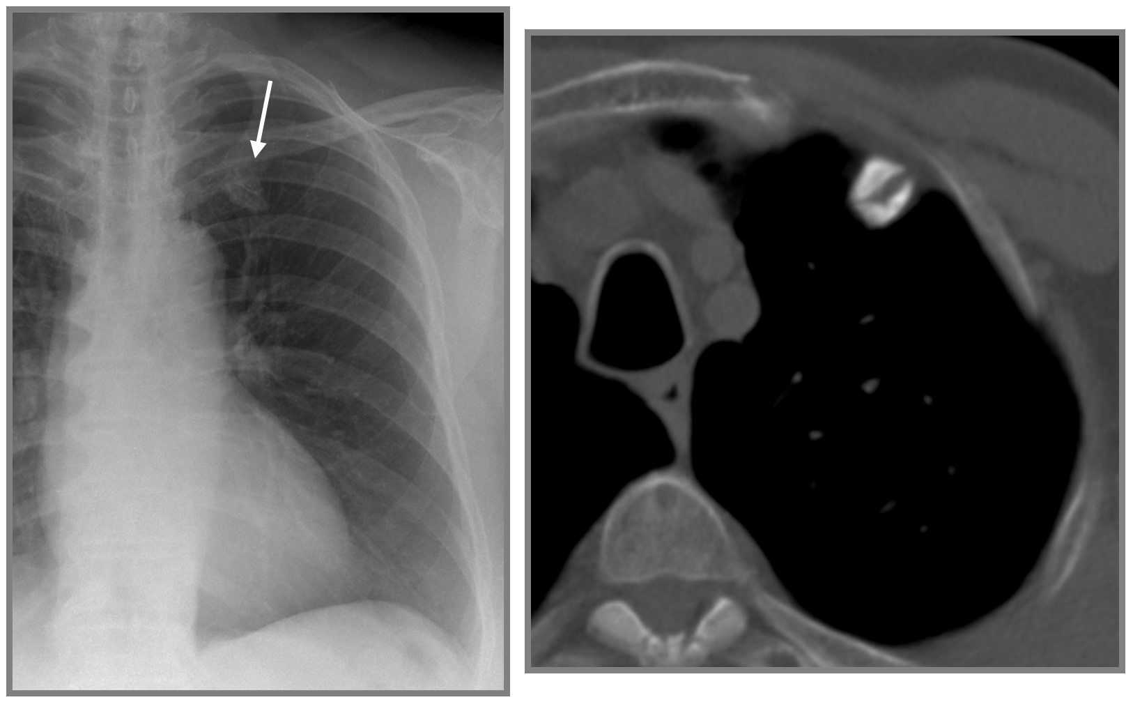
The lateral chest radiograph ( well seen by some of you) shows a nodule overlying the spine ( white arrow). In view of the patient´s history of smoking for many years, a suspicion of possible lung cancer was raised. In this case, without previous images, a chest CT is the procedure of choice.
The CT did not show any pulmonary nodules/masses. But a large osteophyte in the right costovertebral joint at T5 was identified.
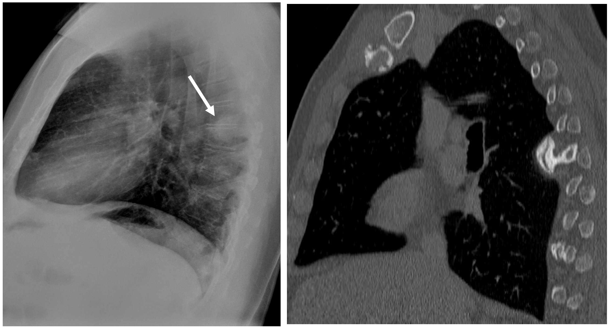
This is not an uncommon cause of false pulmonary nodules, especially in the lateral projection
Osteophytes are so common and variable that may often confuse us. Remember to look at previous images if available and pay attention to the spine and other skeletal structures.
False pulmonary nodule (white arrow) due to osteophytes in the first costo-chondral junction.