José Vilar and Friends Case 47 (Update: Solution!)
Hello everybody.
Here is a new case from my collection to test your ability to see and your knowledge-
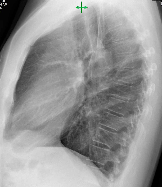
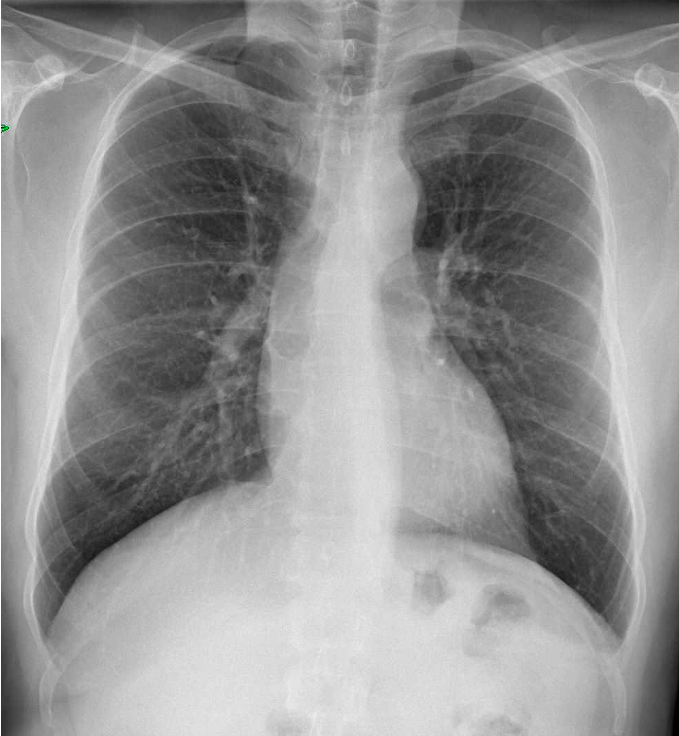
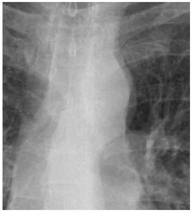
Preoperatory radiographs. No significant symptoms.
As in previous cases three questions:
- Where is the abnormality?
- What could it be?
- What would you do?
Click here for the answer
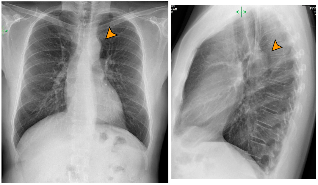
The PA projection shows a double density at the aortic Arch and the lateral an abnormal contour of the aorta behind the trachea.
These images suggest several possibilities:
- Middle mediastinal pathology such as tumours including esophageal, and especially adenopathy.
- Abnormality of the aortic wall or the morphology of the aorta.
As most of you suggested a contrast CT is necessary to make a proper diagnosis.
The contrast CT showed a kink in the aorta at the level of the ligamentum arteriosum (arrows), but no signs of coarctation or mediastinal tumour.
This finding is the typical sign of Pseudocoarctation as Dr Doukopoulou mentioned.
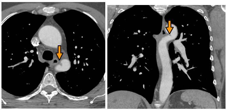
Pseudocoarctation of the aorta is an isolated anomaly that consist of a “twist of the descending aorta at the level of the ligamentum arteriosum. It is not associated with gradient differences in intra-aortic pressure and is usually asymptomatic. Rarely aneurysmal dilatation has been reported and also rarely cardiac anomalies have been found. This entity is not treated, but may give rise to some confusion, especially with aortic coarctation where there is associated symptomatology, pressure gradients, collateral circulation and significant changes of caliber in the aorta before and after the point of stenosis.
Aside from the classical kink of the aorta other findings may be associated such as a high aortic knob above the clavicle.
Points to take away:
- Look at the aortic contour. Mediastinal pathology may be hiding there, but always think first about the vessels. Solve it with a contrast CT.
- Gain the lateral radiograph helps locating the pathology.


A the PA view it is behind the aortic arch, at the laterial view it is posterior at the Th2/Th3 level. I would say it could be schwannoma, enhanced CT should be performed.
At the PA view appears like a double density sign of the aortic knob. At the lateral view there is an incision at the aortic arch.
Possible differential diagnosis coarctation of aorta (despite the lack of ‘figure of 3’ sign) or pseudocoarctation.
I would suggest as next step a contrast enhanced CT.
On L-L projection there is an opacity near aortic arch with image of double contour in P-A projection.
I propose a CT examination to define better this image.
x chest pa view shows dual contour of aortic knuckle and are continuos with the descending aortic line, overlying the aorto pulmonary angle or notch, with distal constriction of aorta.
lateral view suggest similar dual contour of descending aorta just below aortic arch.
suggesting the possibility ductus arteriosus pathology-aneurysm of ductus arteriosus or persistent ductus.
? thinning of posterio inferior border of 4 th and 5th ribs.
PSEUDO COARCTATION
COMMON DIAGNOSIS IS COMMON. I THOUGHT OF WILD IMAGINATION LIKE DUCTAL ANEURYSM.
THANK YOU FOR YOUR ANSWER