José Vilar and Friends Case 54 (Update: Solution and Teaching tips!)
Dear friends, after a short trip to the abdomen, we return to the chest.
Here is a case to prove your ability to detect and diagnose.
This is an 82-year-old man with an important weight loss.
There are several relevant radiological findings in both projections. Can you list them?
Update, Monday 25th October:
Heading to the solution…but not yet!
This has been a strange case. For some reason, and according to the blog counter, more that 14.000 views of the case were registered. My conclusion is that, either the case is too easy or too complicated. Perhaps we are dealing with an adieu to the chest radiograph…
I refuse to accept it, therefore, instead of giving you the answer, let me prolong one more week the case and give you the opportunity to find the solution.
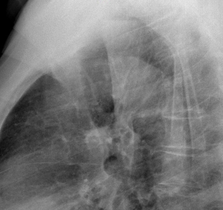
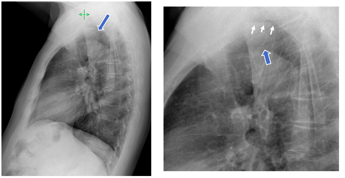
- Here is a close up of the lateral chest radiograph: Findings?

- According to the findings to what specialist would you refer this patient:
- Cardiology
- Pulmonology
- Gastroenterology
Click here for the answer
Solution:
Dear friends,
I have not had any feedback from you until the end, after I showed a close view of the lateral chest radiograph. Then, most of you were right on the spot. Congratulations!
Findings:
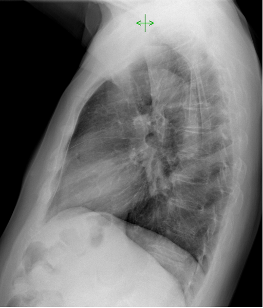
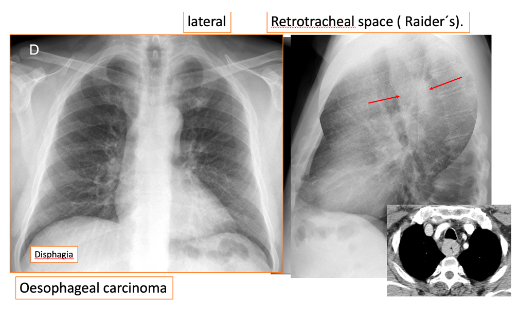
Lateral Chest Radiograph:
- The retro tracheal space ( Raiders space) is occupied ( Blue solid arrow)
- Air fluid level in the upper retro tracheal area ( Small yellow arrows)
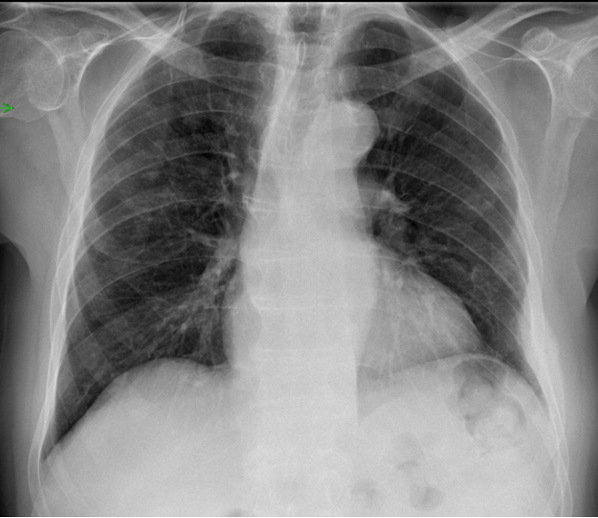
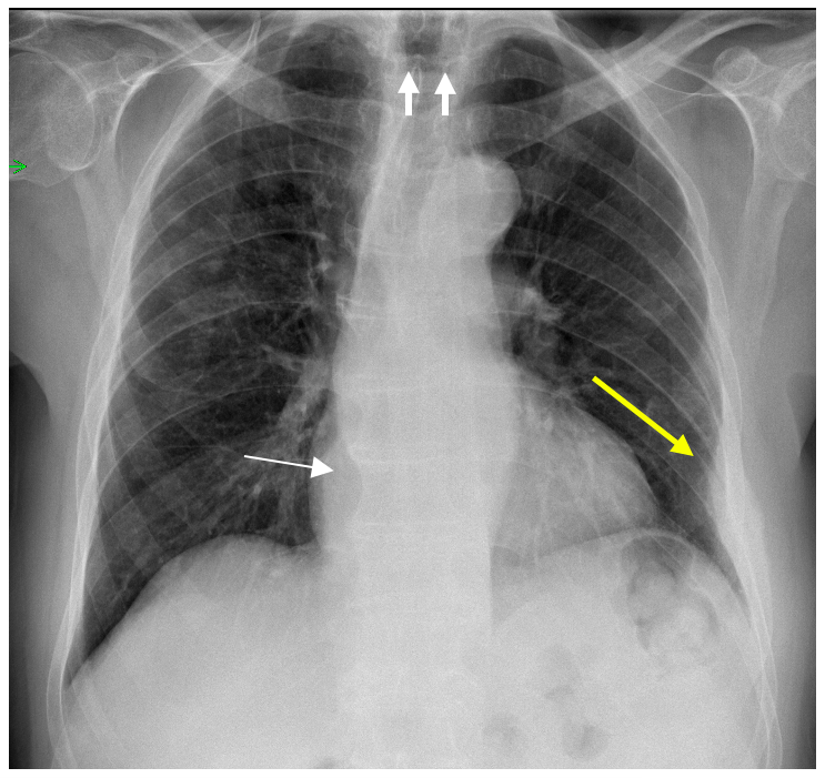
PA Chest Radiograph: The following findings are seen:
- Right upper lobe pulmonary nodules
- The azigo-oesophageal line is displaced outwards. (Arrows)
- Air fluid level above the clavicle.
- Extrapleural lesion in the left hemithorax with possible rib involvement
( Yellow arrow)
Summary of findings in Chest Radiographic examination:
Mediastinal lesion in the region of the oesophagus
Pulmonary nodules
Possible rib lesion
All the findings point to a tumour in the oesophagus.
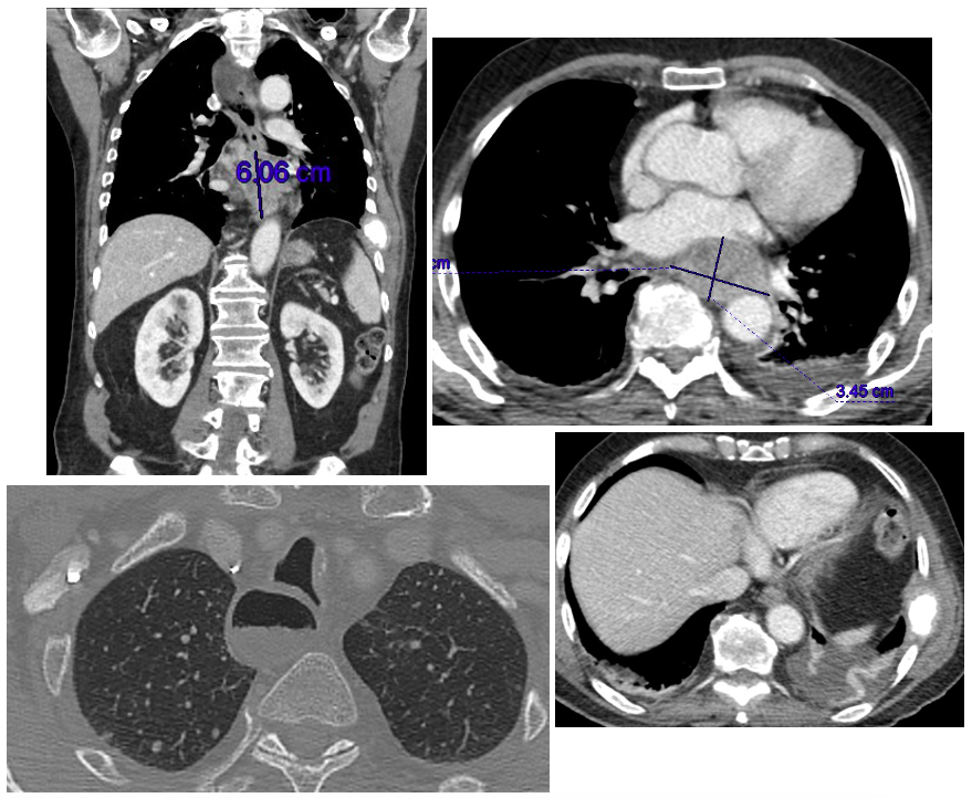
A contrast CT was performed:
1- Oesophagus mass, dilatation of the proximal oesophagus, pulmonary nodules and rib metastases.
Comments:
This case illustrates once again the value of a chest radiographic examination, and especially the lateral projection.
Remember that the in lateral chest radiograph you must look, especially, at three clear spaces, and the spine.
- Retrosternal clear space: Remember the cases of anterior mediastinal masses I have showed previously (Case 39).
- The retro tracheal space or Raider’s space
- The retrocardiac space
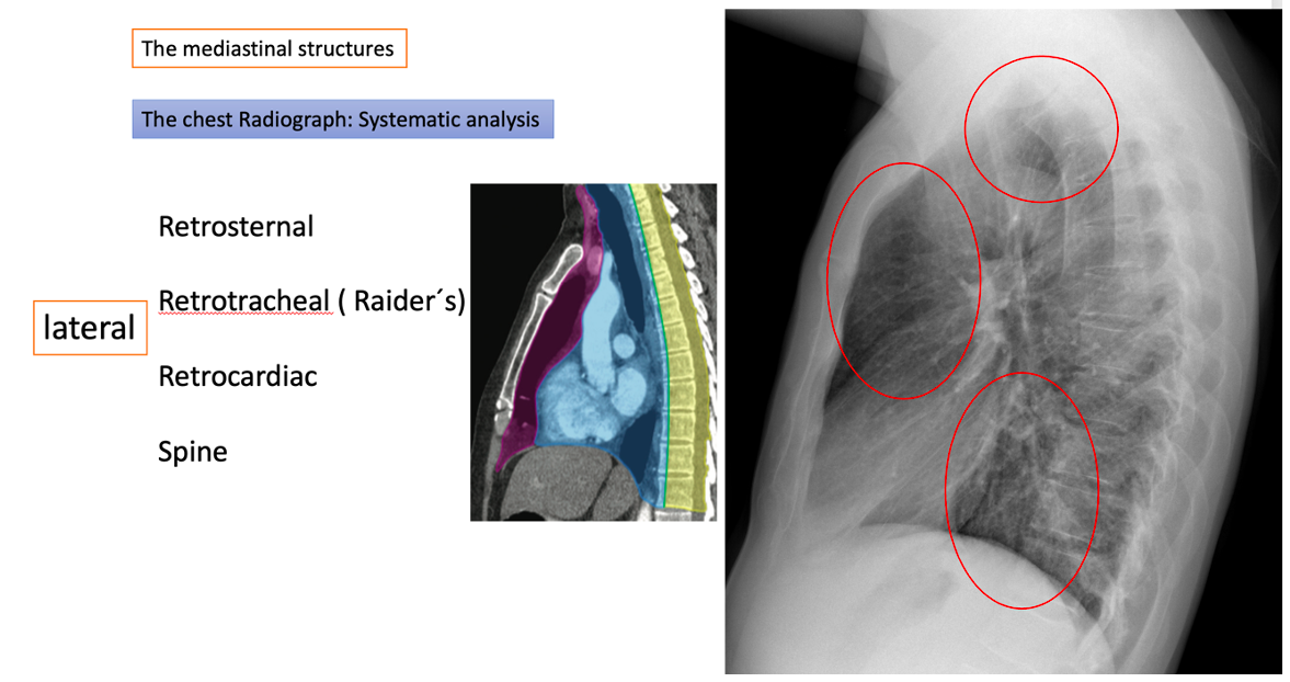
The Raider space (Louis Raider 1973), is a virtual space limited by the trachea anteriorly, the thoracic inlet superiorly, the spine posteriorly and the aortic arch inferiorly.
Different mediastinal pathologies may involve this space: Vascular, Thyroid, Oesophagus, infections.
Some examples of Raider’s space occupation
Here is another case of Oesophageal carcinoma
Two other cases of retro tracheal pathology: An abscess from a dental complication and a right aortic arch.
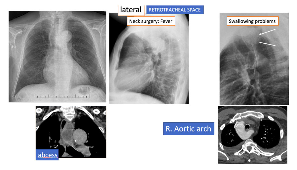
(Slides from J.Vilar. Lecture at ECR 2021)
Tips:
- Look at the lateral radiograph always. Remember the clear spaces.
- Once you have found the pathology…keep looking. In this case a rib fracture may go undetected if you stop looking (Satisfaction of search error).


So many views of this case and no one has so far said a word… Let me give you a hint: look at the mediastinum in both projections
Retrotracheal opacity with an air-fluid level, suggestive of dilated esophagus possibly due to obstruction. Several round nodules in the right upper-middle lung zone. Left lateral 7th rib focal enlargement/deformity with sclerosis and adjacent pleural/extrapleural thickening, suggestive of destructive process or a healing fracture (either simple or pathological).
Overall impression – concerning for esophageal neoplasm with lung and bone metastases.
Dilated oesophagus with air fluid level-secondary achalsia
Multiple lung opacities – metastasis
Multiple enlarged hilar/mediastinal LNs with appreciable dilated gas filled structure retrotracheally presumed to be esophagus . features could be of secondary achlasia with metastatic mediastinal lymphadenopathy
wide mediastinum, air fluid level (in PA and lateral), faint pulmonary nodules (mainly RUL), extrapleural lesion in the left 7th rib.
All together can be aclasia due to distant esophageal tumor with pulmonary and chest wall mets.
Refer to GIT.
Looks like there is opacification of the raider’s triangle, with a suggestion of air-fluid level on both pa and lat. Bilateral pulmonary nodules a left rib lesion. With the hx and your hints, metastatic mid to upper esophageal tumor? Nodules could be mets or maybe just aspiration
¿El señor tiene un parásito alojado en la tráquea? El esófago está dilatado y se ve una opacidad de forma extraña. Y si encima tiene una pérdida de peso importante…
Pensaba que las tenias y ascaris no se veían en radiografía simple. Al gastroenterólogo de cabeza.
Gran caso!!