José Vilar and Friends Case 55 (Update: Solution, Tips and a Goodbye)

Dear all.
Time passes, and I must move on. After several years with the blog “José Vilar and Friends”, more than two million visits and near five hundred thousand visitors, I am finishing my job. It has been a great experience, trying to teach some basic clues, but also learning a lot from you, and from my friends: radiologists from different spots in the world. Fifty-five cases containing some tips to remember, especially (but not exclusively) dealing with plain radiography of the chest. Yes, the chest radiograph, after more than one hundred years, is still alive, but probably mistreated. Fundamental information lays within those images, and I have tried to transmit some clues to help you find it. The most important ones:
- Take your time to read the images (every corner of the image counts)
- Look always at the lateral projection, it contains basic information.
- The previous images, and the clinical scenario may give you the answer.
Basically: DETECT, LOCATE, DIAGNOSE.
But I resist to finish my blog without a last case. This is not a complicated case, rather a simple one, where the three points that I have just mentioned can be applied. It was shown to me a few days ago by Dr Santiago Isarría. He and Dr. Maria Luisa Domingo, from the Thoracic section of the Radiology Department at Hospital Universitario Dr. Peset in Valencia, Spain, have been the support of many of my cases, and a constant inspiration, as has been my dear friend José Caceres from Barcelona, and the many other radiologists that helped me. All of them are fellows who still believe in the value of chest radiology and in the wise use of imaging in the different pathologies that we encounter in our daily practice.
And my gratitude also goes to EBR, ESR ,and the excellent technical help, and editing of Simona Bartovicova and Jabarkhel Kamil.
Here is this last case:
This lady comes for a routine control from the cardiology clinic. She is stable.
Watch carefully: What do you see apart from the cardiomegaly? What would you do?
Update, Wednesday 10, November 2021: More images!
Here are the previous PA chest radiographs
Click here for the answer
Solution:
Dear friends,
This is my last case. As I indicated in the presentation of this case, it has been a pleasure to participate in this blog, and to share with so many people good moments.
As I told you, this is not an exceptional case, yet it contains the basic elements of my fundamental message reading images.
Let us see…

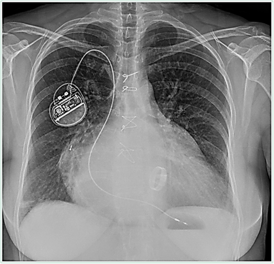
In the initial images there are obvious changes related to cardiac pathology with cardiomegaly, especially a large left atrium and left ventricle. There is a single chamber pacemaker implanted through the right side, and a mitral valve prosthesis.
A small 2 cm metallic object is seen in the PA projection overlying the right heart (Red circle).
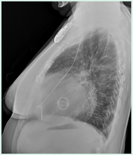
The lateral projection shows that this object has a posterior location, and thus it cannot be in the heart. (The right heart chambers are located anteriorly).
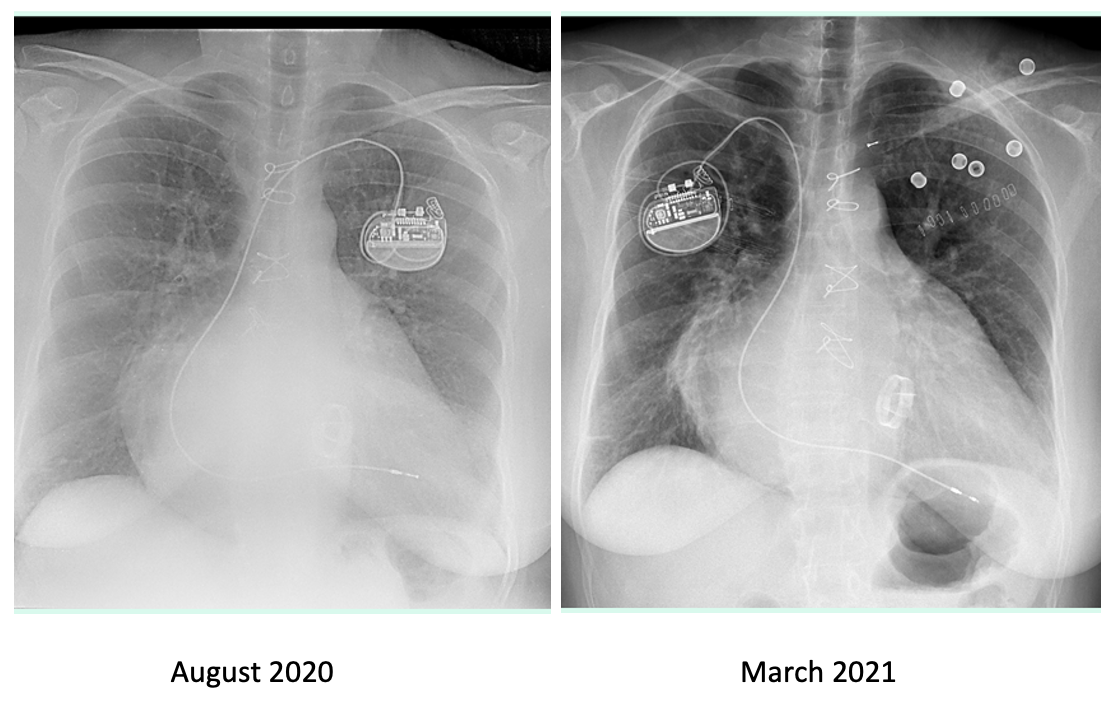
As we can see, the patient had a pacemaker implanted through the left side in 2020.
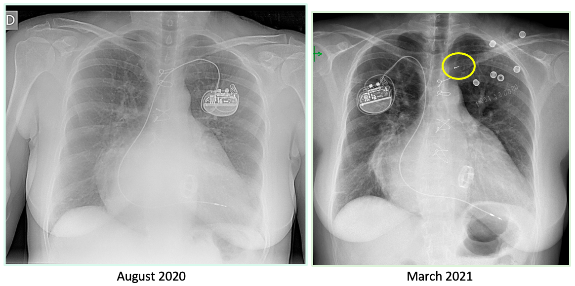
And, in a control obtained in March 2021 when the pacemaker was replaced and implanted via the right side, the metallic object is seen overlying the left clavicle (Yellow circle).
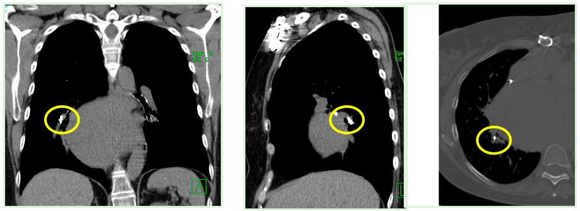
Diagnosis: The logic of the case is that it must be a lead of the electro catheter that was left in the left subclavian vein during the retrieval of the pacemaker in 2020. This metallic lead migrated (“embolized”) to a branch of the right pulmonary artery.
This location can be well seen in an actual non-contrast CT. (Yellow circles)
Rupture of the pacemaker leads may occur, and we must be aware of this complication and report it to the cardiologist.
Tips: This case, as I told, reveals the importance of:
- Looking at the previous studies
- The lateral projection always helps.
- Radiologist reading cardiology radiographs must know the different devices that can be implanted and their possible complications.
I leave you with a reference from our group that could help you, when you are dealing with pacemakers and other implantable devices in chest radiographs.

And this is all. My best wishes to everybody and thank you again for letting me enjoy this blog with all of you. Yet there are many things to undertake.
Two roads diverged in a yellow wood,
And sorry I could not travel both… (R. Frost)

Meanwhile the orange harvest comes to us with renewed promises. A positive premonition…


Realmente vi las imágenes por mucho tiempo y lo único que veo es un clip metálico que en la Rx actual está en relación a la arteria pulmonar derecha y en la previa estaba en campo superior izquierdo… Pero no entiendo bien la relación que tiene.
¡Muy bien Marcos!.Now one has to find out where is that “clip” and how could this object have arrived there. The information is in the images.