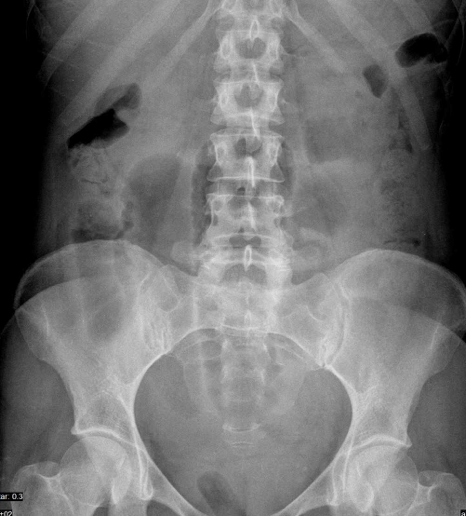
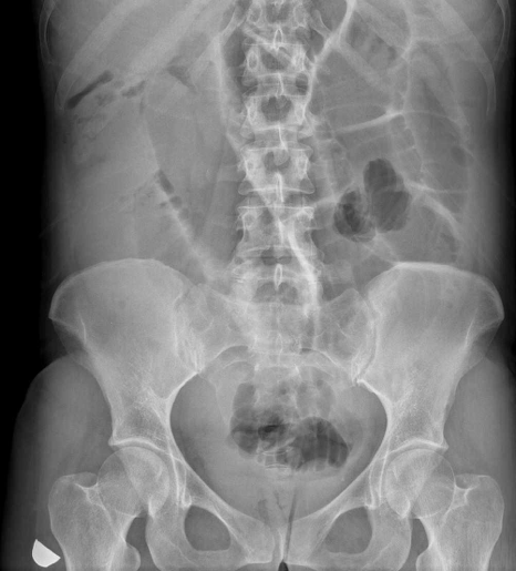
this time we are going to move down into the abdomen. This case was shown to me by my good friend Dr José Vizuete (Hospital Universitario Dr Peset, Valencia) and I think that it shows how we should look at images and use all the information included in them.
This is a 39-year-old woman who presents at the Emergency Room with abdominal pain in two occasions.
Case provided by José Vizuete MD. Hospital Universitario Dr. Peset. Valencia. Spain
Solution: Both abdominal radiographs show dilatation of small bowel loops suggesting a small intestine obstruction. There is a subtle finding in both radiographs taken sometime apart: in the lower pelvis (red arrow) you can see the presence of a Tampon. This means that both examinations were obtained during the menstrual period. In the absence of previous surgery, sings of appendicitis or oncological history, repeated abdominal symptoms coinciding with menses should raise the possibility of Endometriosis.
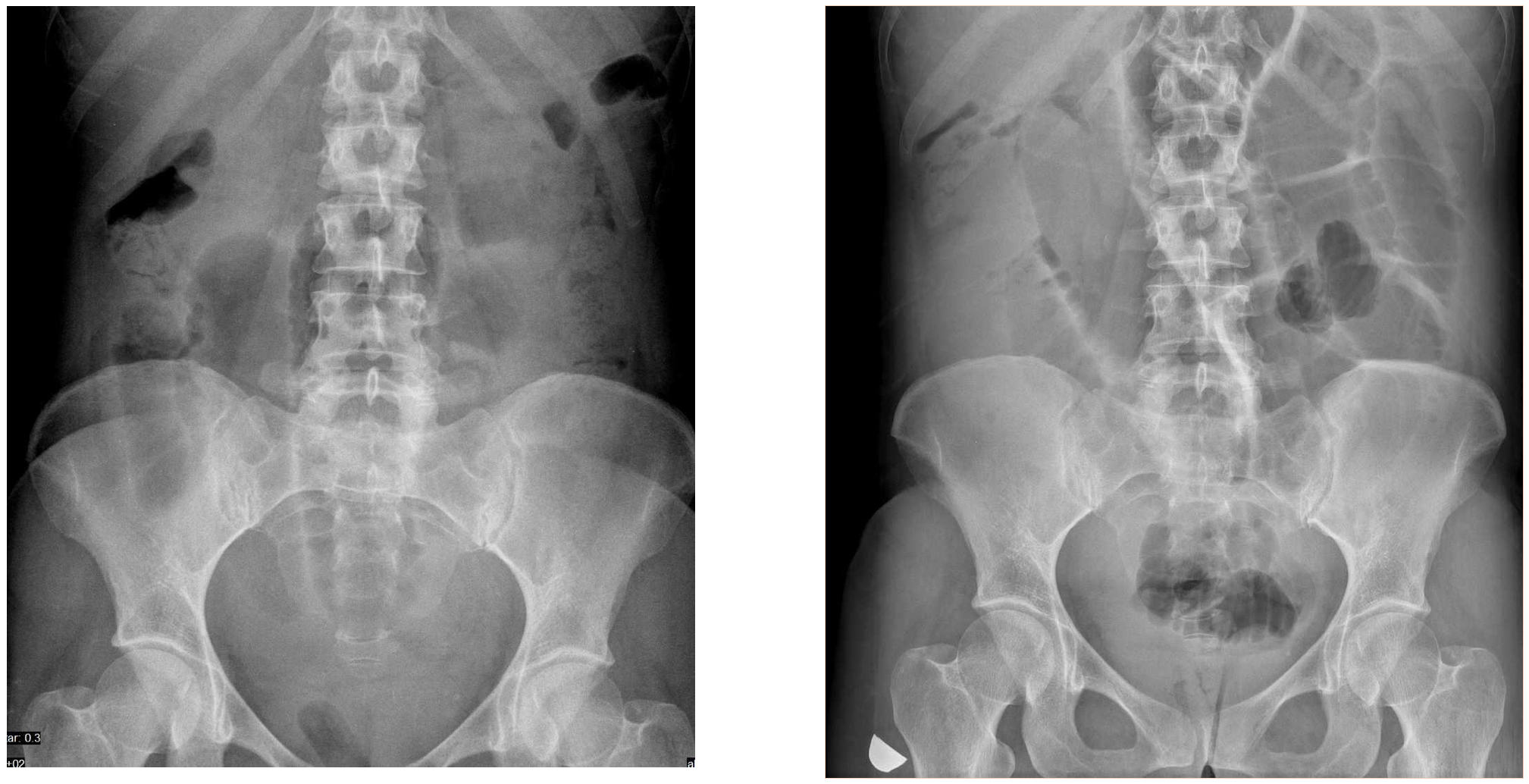
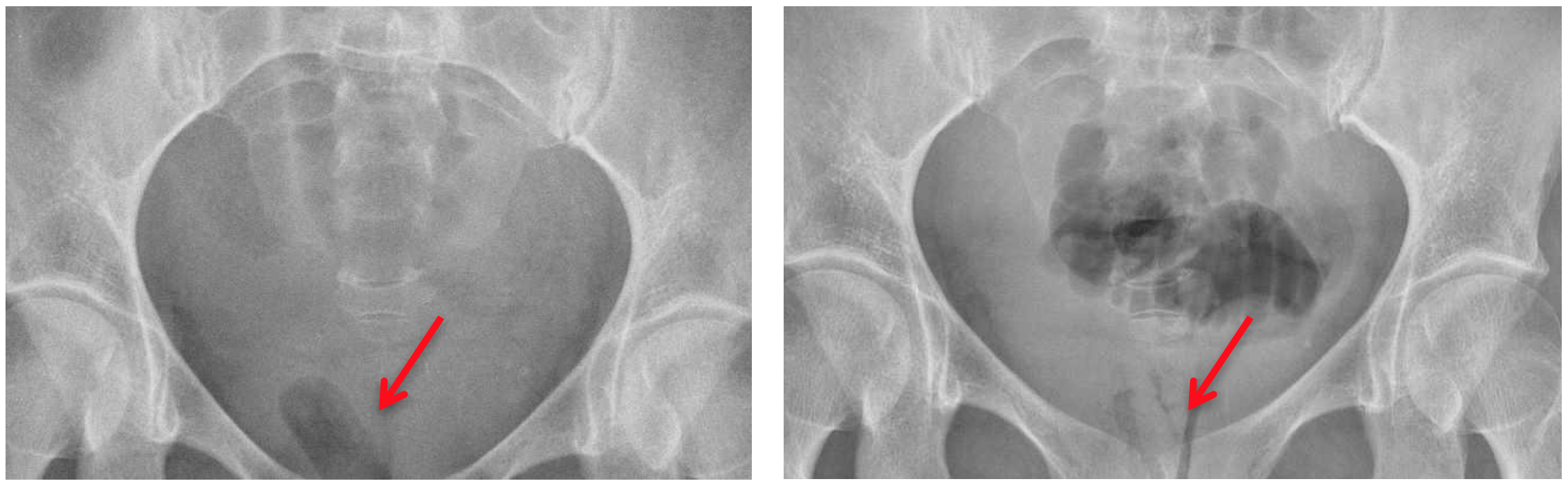
Red arrow shows tampon in both studies providing important clinical information: the symptoms occurred during menses
An ultrasonographic study of the bowel demonstrated the presence of hypoecoic nodules in the bowel serosa (red arrow) a typical finding of this disease.

US examination of the ileocoecal region. A hypoecoic nodule is seen in the outer layer (serosa) of the ileum (red arrow) and the rest of the layers are preserved (white arrow). The bowel loop is distended (ASA PRE) proximal to the Endometriosis nodule (E) and normal distally (ASA POST).
Diagnosis: INTESTINAL ENDOMETRIOSIS
Endometriosis may involve extra uterine structures, mainly around the pelvis, but also I other areas such as the bowel or even the pleura. The endometrial lesions are hypoecoic in ultrasound. In MRI these lesions are hyper intense in T1 and hypo intense in T2 weighted images
Teaching points:
- Look at all the “corners” of the image and use any information present.
- Ultrasound may be useful in the diagnosis of intestinal endometriosis but requires and important level of expertise in the technique of bowel ultrasonography.