Hello friends,
this is a case of a patient with chronic disease.
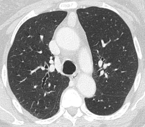
I am showing you two CT images and, as usual, hope to read your comments.
Click here for the answer
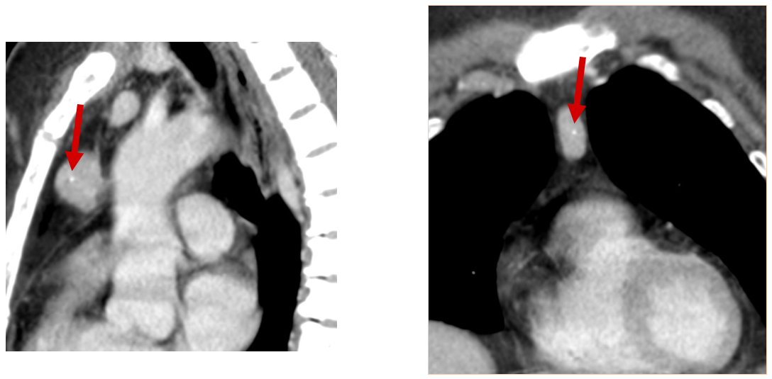
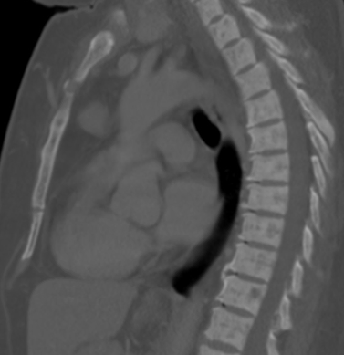
Comment: The CT shows an anterior mediastinal mass/nodule that enhances almost as much as the aorta. The mass has a central calcification (red arrow). In this case I have included a sagittal CT image with bone window settings that shows diffuse bony changes in the spine produced by Hyperparathyroidism. This is the clue to the case.
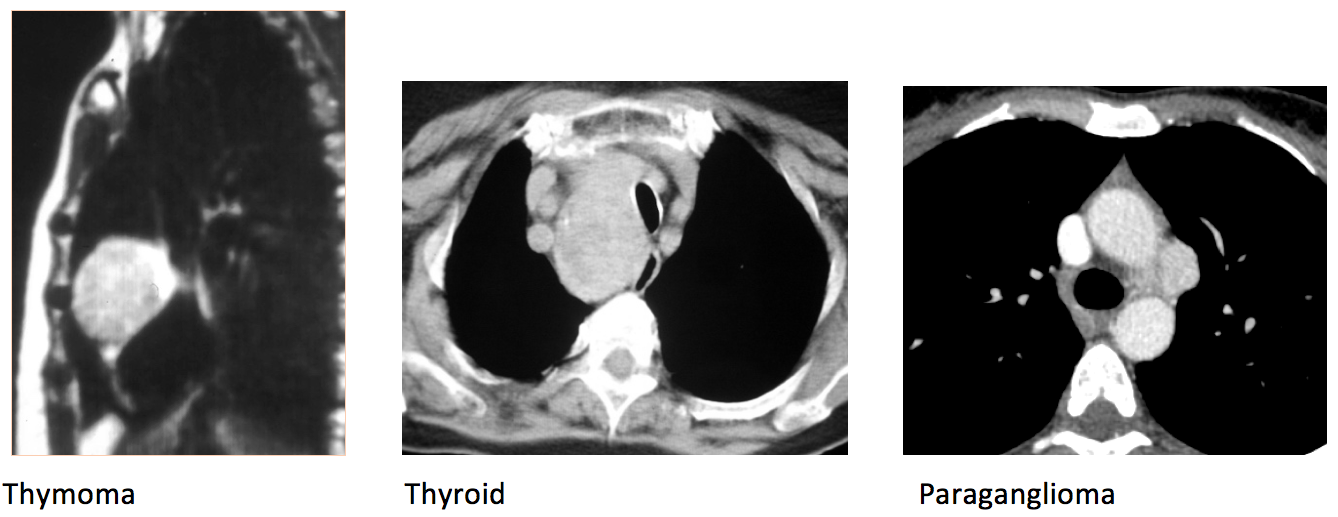
Hypervascular mediastinal masses are most commonly Thymomas, but other causes are adenopathies from hypervascular tumours, Thyroid tumours, Castleman disease, and paragangliomas.
Solution: Primary Hyperparathyroidism with Ectopic parathyroid adenoma
Teaching points:
- Always look for additional findings that may give you the clue in the diagnosis
- Remember that high contrast uptake may help in the differential diagnosis of mediastinal tumours.


