
Dr. Ramiro Hernandez
Hello my Friends.
Today I am presenting you with a case from my good friend Dr. Ramiro Hernandez from Ann Arbor University, Michigan.
He is a well-known pediatric radiologist and, as me, likes to extract good and useful information from plain films.
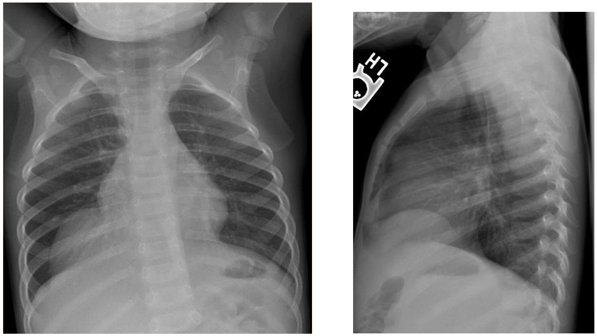
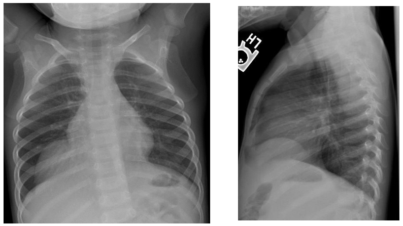
These radiographs belong to a one-year old child with suspected respiratory infection.
Let us see what you think…
Additional information:
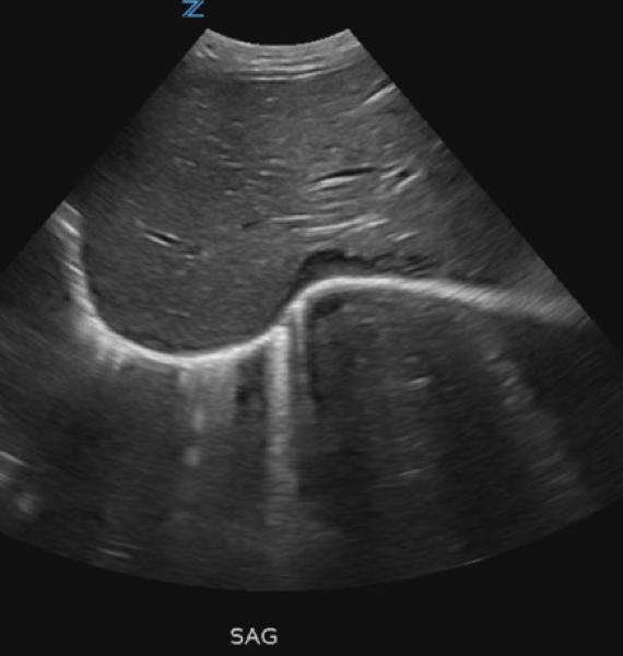
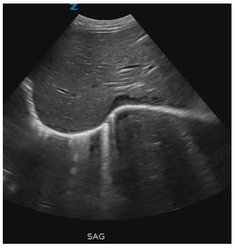
After reading your smart comments, here is an ultrasound image. With this, you must make the diagnosis hopefully.
This time, come back on Monday to find the solution!
Click here for the answer
This is a one-year old child with suspected respiratory infection.
AP and lateral radiographs of the chest and upper abdomen. There is a focal smooth convexity in the anterior and medial right hemidiapragm. The smooth border indicates its extrapleural location.
A differential diagnosis could be established with a hernia ( Morgagni) or a liver mass.
Sagital Ultrasound: The study confirms that there is a deformity of the diaphragm with the normal liver occupying this space. This study excludes liver mass or hernia.
Diagnosis: Congenital eventration of the diaphragm.
Comment: Eventration is defined as a local thinning of the diaphragm causing a bulge. The most common location is the right anteromedial aspect. Eventration is usually focal, thus only part of the diaphragm is elevated. Most eventrations are congenital, but they may also be acquired. The differential diagnosis is with paralysis of the diaphragm that is not focal, hernia, mediastinal masses or cysts and abdominal masses. A pitfall may be a normal exhalation that may mimic a diaphragmatic elevation. Ultrasound usually will solve these diagnostic problems.
Eventration needs treatment only when it produces symptoms.
Teaching point: Think of eventration when a focal bulge of the diaphragm is present.
Two references from Radiographics if you want to know more…
Multimodality Imaging of the Pediatric Diaphragm: Anatomy and Pathologic Conditions
RadioGraphics 2010
Volume: 30Issue: 7pp. 1797-1817
Imaging of the Diaphragm: Anatomy and Function
RadioGraphics 2012
Volume: 32Issue: 2pp. E51-E70
I must congratulate again the colleagues that made a correct diagnosis.


Morgagni’s hernie DD pericardial fat pad
Hello,
There is an abnormality of the diaphragmatic contour on the right medially, partially silhouetting the right heart border. It is well defined and has rounded contours. It likely represents a lesion which is extrapulmonary and could be arising from pleura, diaphragm or from underneath the diaphragm. Differential is broad and I would further investigate with an ultrasound in the first instance.
It could represent a pleural neoplasm (pleuroblastoma?) or loculated effusion (less likely). It could also represent an abnormality of the diaphragm (defect with herniating liver?) or a liver tumor pushing the diaphragm upwards. Other differentials would be pericardial cyst, sequestration, congenital (foregut duplication) and acquired (hydatid) cysts.
Cheers!
Partial eventration of diaphrgm with high liver.
Well-circumscribed mass adjacent to right cardiac border with cardiac silhouette
DD:
*Pericardial cyst
*Morgagnia hernia
*Pericardial fat pad
11 pairs of ribs
Eventration of diaphragm
Mediastinum appears narrow. Status of thymus ?
It looks like Morgagni hernia from US
Eventration of liver right hemithorax.
Eventration of right hemi diaphragm with liver herniation into right hemithorax.
cardiac rhabdomyoma? you cannot see the border of the left atrium on the xray and i think i can see the pericardiac fat shadow/border on the right at the frontal view. on u/s it looks solid- like and eterogenous (could be an amartoma)or maybe breast milk. i think it also correlates with the clinical context awaiting for the answer… thank you
EVENTRACION FOCAL ANTERIOR DEL HEMIDIAFRAGMA DERECHO