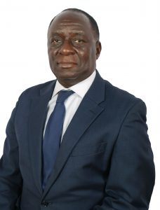
Spotlight on radiology in Uganda
Michael Grace Kawooya is a Professor of Radiology at the Ernest Cook Ultrasound Research and Education Institute and Professor Emeritus at the Makerere University College of Health Sciences in Kampala, Uganda. He has done much for the development of radiology in his country and the rest of Africa, but says efforts must continue to increase the number of radiologists and range of equipment, and to raise awareness of radiation safety. Kawooya believes Africa can learn a lot from European advances. His contributions to improving bilateral cooperation will be rewarded today as he receives ESR Honorary Membership.

Professor Michael G. Kawooya’s contributions to improving bilateral cooperation between Africa and Europe will be rewarded today as he receives ESR Honorary Membership.
ECR Today: How much has radiology advanced in Uganda?
Michael Grace Kawooya: Back in the late 1980s, radiology was very new to medical practice in Uganda, and its contribution to healthcare was not well understood. It was not given priority and was underfunded. There were only two radiologists in the country when I finished my residency and we were overwhelmed with work. Doctors were not willing to undertake radiology residency, fearing that radiologists didn’t earn much. Equipment was scanty and often malfunctioning. Many of these challenges still exist today, but to a lesser extent. Radiology is better understood and its role is now evident. The number or radiologists has increased to almost 70. This year alone, 20 doctors took up radiology residency. The number and range of equipment has also increased.
ECRT: Are there any regional trends in radiology in Africa?
MGK: The same challenges facing radiology in Uganda bedevil most of Africa, but North Africa, which is largely Arabic, and South Africa, which is wealthier, face fewer challenges. In these parts of Africa, radiology has flourished more compared to Central, East, and West Africa. The radiologist-to-population ratio is approximately 1:67,000 in Egypt, 1:1,600,000 in Uganda and 1: 8,000,000 in Malawi. The more affluent regions have higher numbers and range as well as sophistication of imaging equipment. They have more radiology training institutions and undertake more research.
ECRT: Your research interest is ultrasound. Which new developments do you use in daily practice?
MGK: In Uganda, we have embarked on some of these developments, especially in the application of ultrasound in non-traditional areas like anaesthesia, point of care, emergency medicine, musculoskeletal and nerves, oncology and interventional radiology.
Other new, interesting developments include the application of contrast agents elastography, machine learning and artificial intelligence to ultrasound.
ECRT: What has been your experience with ultrasound in a low-resource setting?
MGK: Diffusion of ultrasound in this setting has been explosive. This may be due to the fact that we are starved of high-tech cross-sectional imaging like CT and MRI, and ultrasound has been termed by others as the ‘poor man’s CT and MRI’. In Uganda, one finds an ultrasound machine in almost every corner of the small and major towns and cities. It is feared that many of these machines are manned by untrained practitioners, but in fact many are manned by sonographers. In sub-Saharan Africa, Uganda can boast of having the highest throughput of sonographers from our training institutions. Sadly, some of these Ugandan ultrasound graduates have been ‘brain-drained’ to Europe and other regions. Ultrasound has been maximally exploited in investigation of all parts of the body including brain, chest, cardiovascular system, musculoskeletal and others, whereas in developed countries one may use CT or MRI.
ECRT: What about the use of ultrasound in imaging tropical diseases?
MGK: In diseases like bilharzia, ultrasound plays a key role in epidemiological studies and is applicable in the diagnosis and ultrasound-guided therapy of hydatid disease (echinococcus), which consists of aspiration of the liver cyst and alcohol injection to kill the parasites.
In HIV/AIDS, ultrasound is valuable in demonstrating abdominal morbidity and co-morbidity, especially tuberculosis, and is used to guide diagnostic procedures for obtaining tissue specimens, cells and exudates.
The disease burden for some of these conditions is high and outweighs available resources equipment and trained manpower. Resources for purchasing diagnostic and treatment kits are scarce. Machines may be old with poor resolution. Patients living in remote areas can’t access or afford the much-needed services.
ECRT: You helped found AFROSAFE.rad, a campaign for radiation safety in Africa, a few years ago. What are the first results?
MGK: One major outcome is increased radiation safety awareness by radiation workers, medical workers and the public. There is a rising interest in the formulation of dose or diagnostic reference levels (DRLs) and the use of clinical referral guidelines. There is increased research in radiation protection. Collaboration and networking among African countries and with international organisations and campaign-platforms is growing. The cause of these trends could also be multifactorial and not only AFROSAFE.rad.
ECRT: How is artificial intelligence being implemented in radiological practice in Africa?
MGK: There is a budding interest in artificial intelligence (AI). In Uganda, we are fortunate to have an artificial intelligence laboratory in the Main University of Makerere and one student attempted a study on AI and breast cancer. Our ultrasound training institute ECUREI has written up and submitted a proposal on AI in prostate cancer to an international funder. One major international search engine has shown keen interest in supporting the institute in undertaking projects in AI beneficial to Africa and we are writing up concepts in this regard.
ECRT: What is your relationship with European radiology and the ECR?
MGK: European radiology is very advanced in all areas. Much of what is done in Europe is applicable to Africa and we are glad to learn how to adopt and adapt European advances.
My first experience with the ECR was in 1993 when I first attended the meeting. Several of my colleagues during my fellowship at the Eberhard Karls University in Tübingen, Germany, encouraged me to attend. At the end of the congress, as I boarded a plane to Brussels in which the then ESR President, Prof Albert L Baert, also sat, I said to myself we should also form a similar society and congress in my country and in Africa. The idea of USOFARI and PACORI, the Ugandan and African societies and congresses of radiology, began to form in my mind. As soon as I arrived in Uganda, I got together with colleagues and we decided to create these societies.
Thereafter, I attended the ECR for a few years and then ceased until 2013. Ever since then, I have attended the meeting every year. I like the congress because I acquire new knowledge and improve my skills. I present at least one EPOS poster a year and that motivates me. The EuroSafe Campaign, which I became familiar with at the ECR, has been very inspiring and motivating, and was key in founding AFROSAFE.rad.
I enjoy catching up with old friends and making new friends. This coming year will be a very exciting year. I have always asked myself when the ESR would meet Africa, and it is happening this year! Africa can share its radiology challenges, victories, joys, culture and dreams with the rest of the world, further cementing our friendship and collaboration. And of course I eagerly anticipate receiving the highly esteemed ESR Honorary Membership.


