
Dear Friends
This is the last case of the first semester. It has been a hard year and I need some quiet time. Will see you again on Monday, September 5 with new cases. Relax and enjoy your vacation!
This week’s case follows the pattern of a ‘Meet the Examiner’ presentation, with questions and answers similar to a real examination. Take your time before scrolling down for the answer. And no peeking!
The images were obtained during routine CT screening in a 72-year-old man, heavy smoker.
What would be your diagnosis?
1. Carcinoma in LUL and cyst in LLL
2. Carcinoma in LUL and cystic carcinoma in LLL
3. Tuberculosis
4. None of the above
Read more…

Dear Friends,
This week’s case follows the pattern of a “Meet the Examiner” presentation, with questions and answers similar to a real examination. Take your time before scrolling down for the answer. And no peeking!
The chest radiographs belong to a 54-year-old man in treatment for RUL carcinoma.
What do you see?
Read more…

Dear Friends,
This week’s case follows the pattern of a “Meet the examiner” presentation, with questions and answers similar to a real examination. Take your time before scrolling down for the answer. No peeking! My good friend Dr. Jordi Andreu has prepared the case.
Chest radiographs belong to a 69-year-old woman with cough, fever and purulent sputum for the last ten days. History of TB thirty years ago.
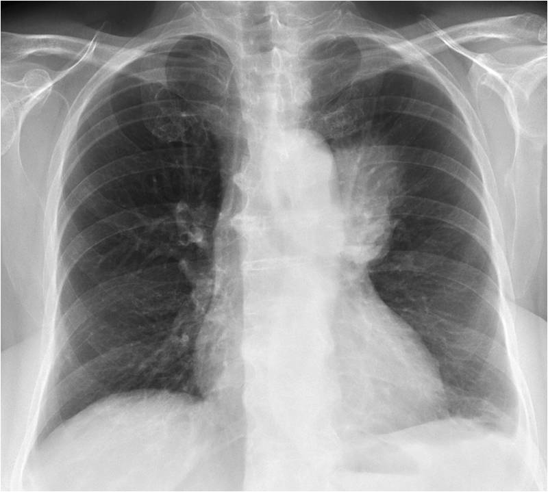
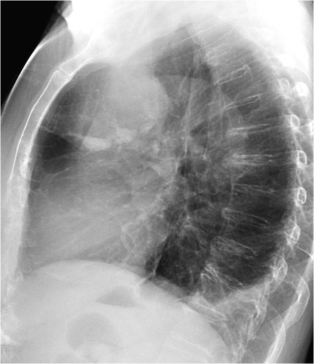
Diagnosis:
1. Reactivation TB
2. Mediastinal cyst
3. Mediastinal tumour
4. None of the above
Read more…
Dear Friends,
This week I’m showing a new ‘Face the Examiner’ case. Presenting the radiographs of a 49-year-old woman with dyspnea.

Dear friends,
This week I’m presenting you another new ‘Face the Examiner’ case which simulates a real examination. Showing radiographs of a 35-year-old male with high fever and left pleuritic pain.
Dear Friends,
As some of you may be taking the Diploma examination during ECR 2013, I’d like to introduce a new type of post that I call ‘Face the Examiner’. Its purpose is to simulate a real examination: images will be shown and you will be asked to describe the findings. You should then offer a differential diagnosis and suggest a procedure that will confirm your preferred option.
The only difference with respect to a real examination is that you will be given the correct answers during the exercise. I’ll try to keep it simple by not giving long lists of possible diagnoses and by making sure the possibilities are coherent with the imaging features.
Our first ‘Face the Examiner’ case concerns preoperative chest radiographs in a 75-year-old man with prostate carcinoma.


