José Vilar and Friends Case 53 (Solution+Teaching tips!)

 Hello everybody.
This time I have had a minor medical problem, and have asked one of my former colleagues, a young radiologist from Hospital Universitario Dr. Peset, in Valencia to help me. Dr José Vizuete is a splendid radiologist, and one of his areas of domain is the abdomen. (He was co-author of our case 3, a case of ganglioneuroblastoma).
So, we will leave the thorax this time and visit our neighbour the abdomen.
This is a 33-year-old man with abdominal pain. Here is an abdominal CT and an MRI.
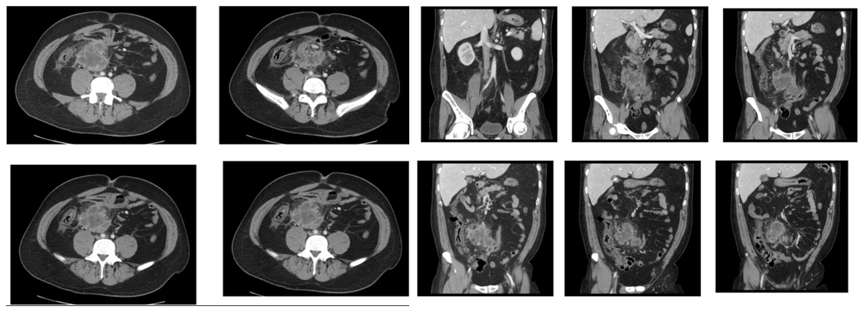
CT
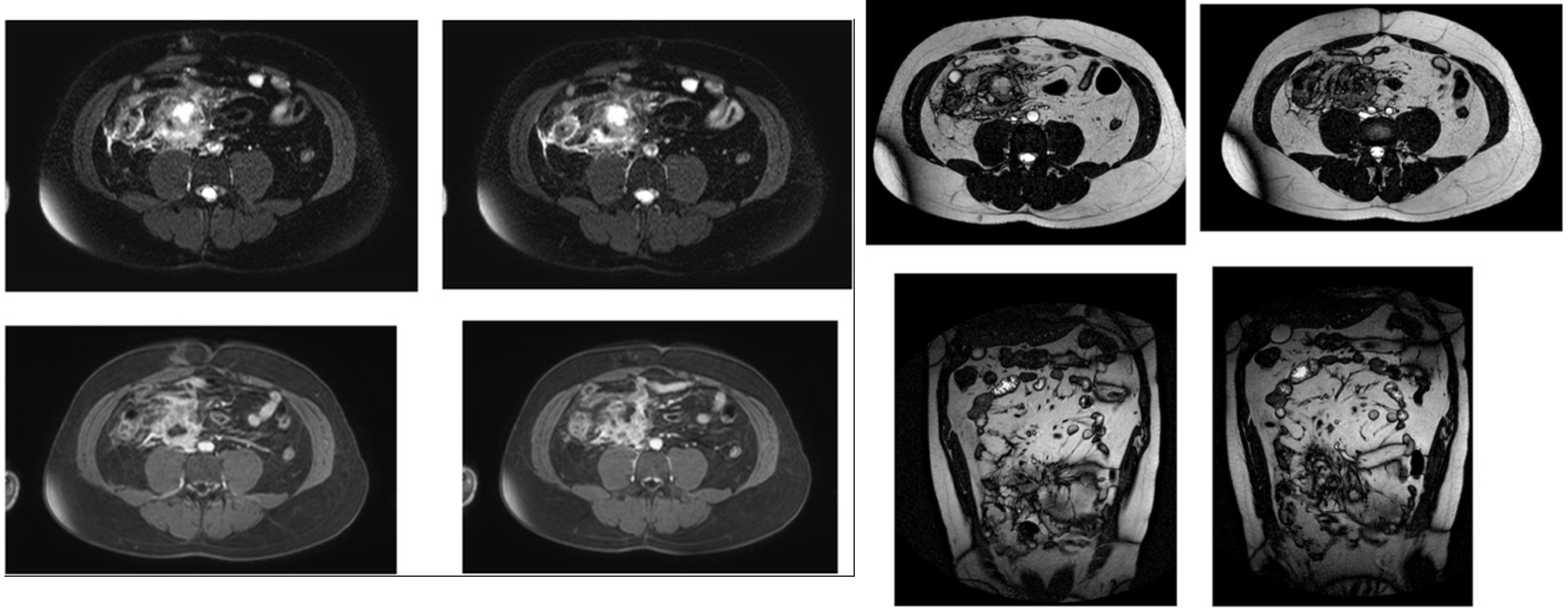
MRI
All Dr Vizuete and I would like is to have a differential diagnosis, but, of course, if you want to suggest a specific pathology feel free to do it. (If you don’t feel safe with the abdomen, you can ask your colleagues, as I usually do). Good luck!
Click here for the answer
Solution:
This has been an exceptional case, and I realise that for a group more interested in the chest its difficult. A challenge that few decided to confront. ( although more than 2000 individuals visited the case), Dr.Marcos Mestas did that effort, and localised the pathology. Well done.
Let me transmit to you the information provided by Dr José Vizuete regarding the explanation of this case.
Findings:
Mass in the root of the mesentery of poorly defined borders. There areas of necrosis and others that enhance with contrast.(Arrows)
In T2 there are hypointense areas that are hyperintense in T2.
The mass does not originate in bowel or other peritoneal or extraperitoneal organs.
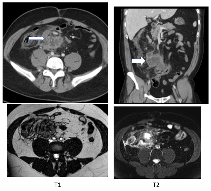
Therefore we are dealing with a Mesenteric mass.
The lesion was biopsied and chronic inflammatory changes were identified. Neoplasm was excluded. It was diagnosed as Retractile Mesenteritis
- Differential Diagnosis of Mesenteric Masses:
- Peritoneal fibromatosis ( Desmoid tumour)
- Carcinoid
- Inflammatory pseudotumour
- Metastases
- Lymphoma
- Retractile mesenteritis
Other names for this entity: Idiopathic panniculitis of the mesentery, mesenteric lipodystrophy, lipogranulomatosis, multifocal sclerosis of the mesentery.
Retractile mesenteritis is defined as an idiopathic process, characterized by mesenteric masses. It is a chronic nonspecific inflammation, along with fat necrosis and fibrosis.
- Imaging:
- CT: Mesenteric masses well or poorly defined with soft tissue and fatty densities. It may involve vessels preserving the fat layer. It may have calcifications and mimic cystic lesions.
- MRI: Low or intermediate signal in T1 ad low signal in T2 although some cases have been described of high T2 signal when there a myxomatous component and fat necrosis.
Tip: Remember the mesentery as a cause of masses in the abdomen. The deep location makes CT and MRI the best imaging tools to analyse mesenteric masses.

RadioGraphics 2003; 23:457– 473


Let me see…Can you locate the pathology?
Se ve una colección multiloculada (absceso) en el mesenterio con estriación de la grasa adyacente. También me parece ver el colon derecho con reemplazo graso en su pared. En un paciente joven pienso en una complicación de una enfermedad inflamatoria intestinal.
Como algo más raro no sé como se ve un divertículo de Meckel complicado pero quizás podría ser un diferencial.
Por último un tumor abscedado aunque me no lo creo. Paciente joven tiene que ser algo inflamatorio infeccioso.