B-0950 Clinical feasibility of contrast-enhanced dual-energy mammography (CEDEM) with a tungsten (W)/titanium (Ti) anode/filter combination: a prototype report
T. Knogler, R. Leithner, M. Hörnig, F. Semturs, M. Waitzbauer, G. Langs, P. Homolka, K. Pinker-Domenig, T.H. Helbich | Monday, March 11, 14:00 – 15:30 / Room F1
Purpose: To test the feasibility of CEDEM with a W/Ti anode/filter combination for high energy images in a clinical setting.
Methods and Materials: Fifteen female patients with 15 breast lesions were included in this study. CEDEM was performed with a Mammomat Inspiration prototype (Siemens, Germany) before and after i.v. administration of 2-ml Iomeron® 400 (Bracco, Italy) per kg b.w. at a rate of 3.5ml/sec. Dual-energy images were acquired with 28-32kVp and a W/Rhodium (Rh) anode/filter combination for low-energy and 49 kVp and a W/Ti anode filter combination for high energy. Weighted subtraction images were computed for diagnostic work-up. Images were assessed by two readers with respect to lesion-enhancement and image quality. A histological work-up was performed in all lesions.
Results: Histopathology revealed eight malignant lesions and seven benign lesions (size range from 8 to 38mm). All malignant lesions enhanced and were seen from both readers on weighted subtraction images. Benign lesions did not enhance thus they were not visualised on weighted subtraction images. Image quality was rated excellent from both readers. Based on the visibility of the lesion CEDEM allowed an accurate differentiation of benign and malignant breast lesions.
Conclusion: CEDEM with a W/Ti anode/filter combination is suitable and feasible. Lesion visibility and image quality were excellent. Further research is needed to determine the value of CEDEM in a clinical setting.

A-585 B. Peripheral skeleton
V.N. Cassar-Pullicino | Monday, March 11, 10:30 – 12:00 / Room A
In this interactive session focussing on peripheral musculoskeletal lesions presenting to the Accident and Emergency Department, we will review the strengths and weaknesses of the imaging modalities applied to chosen bone, joint and soft tissue lesions. The imaging-based approach will strengthen the understanding of the different pathological entities which vary from trauma to infection in both the paediatric and adult age groups.
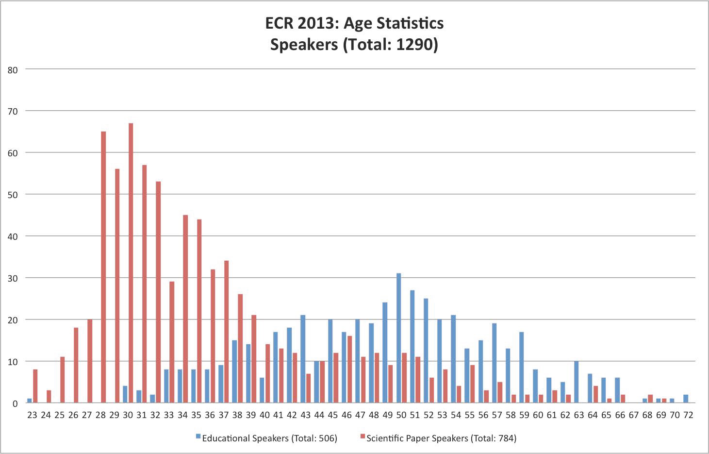
The graph below shows the ages of speakers at ECR 2013. While the most common age of scientific paper presenters was 30, the most common age among educational speakers was 50.
Your ECR future is bright: today’s presenters, tomorrow’s teachers …


A-618 C. Cystic fibrosis and other bronchiectatic diseases
M.U. Puderbach | Monday, March 11, 16:00 – 17:30 / Room I/K
Several different diseases go along with the development of bronchiectasis. Bronchiectasis may result from chronic infection, proximal airway obstruction, or congenital bronchial abnormality. In cystic fibrosis, bronchiectasis is one of the key features of lung involvement. Bronchiectasis can present with a variety of non-specific clinical symptoms, including hemoptysis, cough, and hypoxia. Bronchiectasis is defined as localized or diffuse irreversible dilatation of the cartilage-containing airways or bronchi. The imaging gold standard for bronchiectasis is thin-section CT. Morphologic criteria on thin-section CT scans include bronchial dilatation with respect to the accompanying pulmonary artery (signet ring sign), lack of tapering of bronchi, and identification of bronchi within 1 cm of the pleural surface. Bronchiectasis may be classified as cylindric, varicose, or cystic, depending on the appearance of the affected bronchi. It is often accompanied by bronchial wall thickening, mucoid impaction, and small-airways abnormalities. Besides CT, nowadays MRI of the lung is able to image the relevant morphological features of bronchiectasis. In addition, functional changes due to bronchiectasis can be studied.

A-187 A. RF ablation
J. del Cura | Friday, March 8, 16:00 – 17:30 / Room N/O
RF ablation is currently indicated in HCC as curative treatment in Child-Pugh A-B patients with: single <2 cm nodule and not candidates for transplation, 1-3 nodules <3 cm and not candidates for resection or transplantation. RF can be also performed in patients waiting for liver transplantation. Some studies suggest that survival does not differ between RF ablation and resection in 5 cm because of the high possibility of recurrence. Different types of electrodes can be used: internally cooled, cluster, expandable, with saline instillation. Although results can be good with any of them, every type of device requires a different technique of ablation. Obtaining a margin of at least 0.5 cm of ablated tissue around the tumour is key to avoid recurrences. Combined treatments like combining chemoembolization or PEI with RFA can be useful to increase the ablation volume. Published data show a pooled 5-year survival of 48-55%, with better outcomes in Child-Pugh A patients. In candidates for surgery, 5-year survival is similar to resection: 76 %. RFA is safe: major complications appear in 10 % and reported mortality is 0.15%. Tumours located subcapsular or near major vessels, biliary tree or bowel are more prone to complications. Laparoscopic ablation can be an alternative in these cases. Imaging follow‑up with CT, MRI or CEUS is performed to assess the outcome and detect recurrences, new lesions or complications. Although not well established, most protocols include an immediate post-procedure imaging, 1-month follow-up and explorations every 3 or 6 months for 2-3 years.

A-438 A. The current criteria for nodal involvement on CT/MRI
W. Schima | Sunday, March 10, 14:00 – 15:30 / Room E2
In a variety of diseases, such as metastatic disease, lymphoma and inflammation, lymph node enlargement can be seen. Thus, lymph node characterization is important to differentiate between benign and malignant disease. It is based on size (short axis diameter) and morphologic criteria, such as shape, homogeneity, and contrast enhancement. For abdominal nodes, location-specific size criteria apply (upper limit of normal: lower paraaortic 11 mm, upper paraaortic 9 mm, gastrohepatic ligament 8 mm, portocaval space 10 mm, retrocrural space 6 mm; pelvic nodes 10 mm). However, in clinical practice, often a universal size threshold of 10 mm is used in abdominal imaging. In chest CT, an upper limit of normal of 10 mm is universally applied. However, size criteria alone are unreliable: CT for lung cancer staging has a pooled sensitvity of 51 % (i.e., false negative diagnoses of metastatic deposits in normal-sized nodes), and a specificity of 86 % (i.e., false positive diagnoses due to enlarged reactive nodes). With MRI, the same size criteria apply. However, additionally features such as central necrosis (T2w fatsat or gadolinium-enhanced images) are suggestive of metastasis (or suppurative infection). Lymph node-specific USPIO MR agents can depict tumour deposits in subcentimeter pelvic nodes. Unfortunately, they did not reach market approval. DWI is helpful in identifying in lymph nodes as they exhibit high SI with higher b-values. However, diffusion pattern of benign and malignant nodes overlap, so that ADC values do not aid in characterization. Despite the use of modern MDCT and MRI techniques, lymph node characterization needs further improvement.

B-0801 Treatment response assessment in Hodgkin lymphoma: in search for morphological correlates of metabolic activity
T. Knogler, G. Karanikas, M. Weber, K. El-Rabadi, M.E. Mayerhoefer | Monday, March 11, 10:30 – 12:00 / Room F1
Purpose: To predict early treatment response with three-dimensional texture features (TF) in patients with Hodgkin lymphoma (HL) after radio-chemotherapy extracted from contrast-enhanced CT.
Methods and Materials: 21 patients with histologically proven HL were included in this study. Contrast-enhanced (18)F-FDG PET/CT was obtained on a dedicated PET/CT scanner. Volumes-of-interest and long- and short-axis diameter were manually defined on 48 HL manifestations prior and post-radio-chemotherapy on the CT image stack. Three-dimensional texture features derived from the grey-level histogram, co-occurrence matrix, run-length matrix and absolute gradient were calculated for the VOIs. A stepwise logistic regression with forward selection was performed to find classic radiologic features (i.e. lesion diameter, lesion volume) and TF, which correctly classify treatment response (i.e. full response and partial response). Classification in PET/CT was used as reference.
Results: Difference in short axis diameter best fit as classic feature with a sensitivity of 100 %, a specificity of 54,5% and an accuracy of 89,6%. Combination of “S_0_0_1_Entrp” and difference of “vertl_fraction” best fit as TF with a sensitivity of 97,3%, a specificity of 72,7% and an accuracy of 91,7%.
Conclusion: Texture features extracted from contrast-enhanced CT in patients with HL are superior in differentiation between responders and non-responders without the need for PET examinations, compared to classical radiological features. However, PET/CT as state-of-the-art imaging technique has a sensitivity and specificity >90%, so that further research with larger patient number is needed to investigate this new method.

At each ECR since 2008, the ‘ESR meets’ programme has included a partner discipline along with the three guest countries, as a way to build formal bridges between the European Society of Radiology (ESR) and other branches of medicine, and to give congress participants an opportunity to learn about something a little different. At ECR 2014 the programme includes a visit from undoubtedly the largest medical discipline to take part in the initiative so far: cardiology, represented by one of the biggest medical societies in Europe, the European Society of Cardiology (ESC). Cardiology has much in common with radiology, but this is the first time that the two European societies have come together for an official joint session at a major meeting.
Despite some well-known points of controversy between the two disciplines, concerning professional ‘turf’, the overriding message during this afternoon’s ‘ESR meets ESC’ session will be one of cooperation and mutual understanding. Prof. Panos Vardas, president of the European Society of Cardiology, who will co-preside at the session, believes that the blurring of borders between subspecialties makes this kind of exchange of knowledge a must.
Read more…

A-445 Biliary procedures
M. Krokidis, A.A. Hatzidakis | Sunday, March 10, 14:00 – 15:30 / Room F1
Palliative Percutaneous Transhepatic Biliary Drainage (PTBD) is a therapeutic procedure leading to drainage of the obstructed bile duct system. If endoscopy is not possible and if patient is inoperable, then the percutaneous treatment is indicated. Drainage of the bile ducts is performed with a small plastic multiple hole pigtail catheter. Self-locking catheters are preferred in order to minimize the dislocation risk. The percutaneous catheter is pushed through the malignant stricture, so that bile is draining through the catheter towards the bowel loops. Technical success rate of percutaneous biliary drainage can reach nearly 100 % in experienced hands, while the major complications rate is usually lower than 5 %. Clinical efficacy is usually lower, but still over 90 %. The drainage procedure can be extended with the placement of a permanent metallic stent, which keeps the stenosed biliary duct patent, without need for a catheter. Metallic biliary stents have been proved as the best palliative treatment of non-resectable malignant obstructive jaundice, allowing longer patency rates than plastic endoprostheses. The technique is safe, with low-complication rate and procedure-related mortality between 0.8 and 3.4%. Still controversial remains in the timing between initial drainage and metallic stent placement, as well as the question of balloon dilatation before stent insertion. There is evidence that if the initial transhepatic drainage is completed without causing any severe complications, especially bleeding in form of haemobilia, primary metallic stenting can follow as a single-step procedure.

B-0829 Feasibility of abdominal diffusion Kurtosis imaging compared to standard diffusion weighted imaging at 1.5 and 3 Tesla
H. Haubenreisser, J. Hansmann, A. Lemke, J. Wambsganss, S.O. Schönberg, U. Attenberger | Monday, March 11, 10:30 – 12:00 / Room I/K
Purpose: To demonstrate the feasibility of diffusion kurtosis imaging (DKI) for the depiction and differentiation of liver and kidney abnormalities in comparison to standard diffusion weighted imaging (DWI) at 1.5 and 3 T.
Methods and Materials: 109 consecutive patients underwent a routine abdominal MR protocol including standard DWI and DKI. 1.5 and 3 T b-values for DKI were identical: b=0-100-500-1000-1500-2000 s/mm². 55 liver and 36 kidney lesions were evaluated at 1.5 T, and 16 liver and 11 kidney lesions, respectively, at 3 T. DKI was assessed by an in-house built software. Kurtosis values were quantified by region-of-interest analysis and compared between lesions and normal parenchyma by a Wilcoxon rank sum test.
Results: Mean kidney kurtosis values were identical at 1.5 and 3 T in normal parenchyma (K=0.5) and cysts (K=0.4). Differences dependent on field strength were only found in malignant tumours (0.8 vs 0.5). At 1.5 as well as 3 T, cysts, tumours and normal kidney parenchyma could be differentiated with significance (p<0.0413) using the kurtosis values. At both field strengths, cysts, benign and malignant liver tumours could be differentiated with statistical significance from normal parenchyma (p<0.001, p<0.0071, p<0.001). However, malignant and benign liver tumours were significantly different at 1.5 T (p=0.0382). Abnormalities and normal parenchyma could be visually differentiated on both, DWI and DKI.
Conclusion: DKI of kidney and liver abnormalities is feasible at both, 1.5 and 3 T and allows for a quantified differentiation between normal parenchyma and pathologic lesions.


