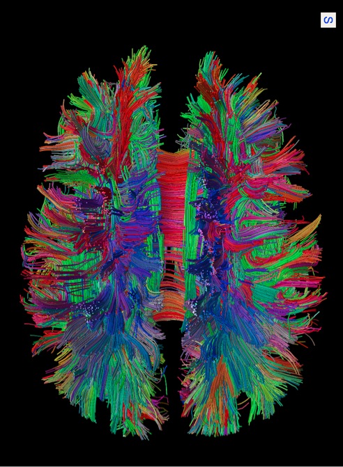
ECR On Demand Preview: The human connectome #NH 7 #A-158

ECROnDemandPreview_Blog_Picture_05

NH 7 – The human connectome, A-158 – Connectomics in brain pathology (M.P. v.d. Heuvel)

A short preview of lecture A-158 ‘Connectomics in brain pathology’, from the session NH 7 ‘The human connectome: a comprehensive map of brain connections’ at ECR 2014, given by M.P. van den Heuvel from Utrecht, Netherlands.

Watch the whole lecture and many more at http://ipp.myESR.org
Direct link: http://bit.ly/The_human_connectome

Friday, March 7, 16:00 – 17:30 / Room Board Room B

Abstract:

Healthy brain function depends on efficient functional communication within a complex network of structural neural connections, a network known as the connectome. Conversely, damage to the brain’s network, disrupting local neuronal processes and/or global communication between remote functional systems may lead to brain dysfunction. In the last few years, emerging evidence from a wide variety of studies suggests that connectome abnormalities may indeed play an important role in the aetiology of several brain disorders. In my talk, I will discuss the results of recent studies suggesting an important role for affected connectome organization in a number of neurological and psychiatric disorders. In particular, I will highlight the findings of affected functional and structural brain network in neurodegenerative disorders such as Alzheimer’s and ALS, as well as discuss how the application of network science and connectomics may aid our understanding of the biological basis of psychiatric disorders such as autism and schizophrenia.

25
Oct 2014
POSTED BY
POSTED IN ,
DISCUSSION 0 Comments

ECR on Demand Preview: The hand and wrist #RC 1910 #A-585

ECROnDemandPreview_Blog_Picture_04

RC 1910 – The hand and wrist, A-585 C- Tumours and tumour-like lesions

A short preview of lecture A-585 ‘C. Tumours and tumour-like lesions’, from the session RC 1910 ‘The hand and wrist’ at ECR 2014, given by E. Llopis from Valencia, Spain.

Watch the whole lecture and many more at http://ipp.myESR.org

Direct link: http://bit.ly/The_hand_and_wrist

Monday, March 10, 16:00 – 17:30 / Room E1

Abstract:

Radiological study of the wrist and hand is challenging due to its complex anatomy with many small structures and the number of normal bone and soft tissue variants that might mimic injuries. Moreover, many findings can be asymptomatic. Their knowledge is important to avoid misdiagnosis. During this lecture we will also review the role of the different imaging modalities, such as plain films for wrist alignment and bone structures as well as the important role of US and MR in differentiating tumour from tumour-like conditions. We will become familiar with some specific radiological findings that allow us to make accurate diagnoses of soft tissue and bone lesions.

11
Oct 2014
POSTED BY
POSTED IN ,
DISCUSSION 0 Comments

ECR on Demand Preview: RECIST criteria #CC718 #A-187

ECROnDemandPreview_Blog_Picture

CC 718 – Imaging after systemic therapies: the standards, A-187 A. RECIST criteria (Y. Menu)

A short preview of lecture CC 718 ‘Imaging after systemic therapies: the standards’, from the session A-187 ‘A. RECIST criteria’ at ECR 2014, given by Y. Menu from Paris, France.

Watch the whole lecture and many more at http://ipp.myESR.org
Direct link: http://bit.ly/Imaging_after_systemic_…


Friday, March 7, 16:00 – 17:30 / Room Conf. Room M3

Abstract:

The routine practice of oncologic imaging requires standardisation, which means that we need to harmonise technical protocols and agree on the meaning of selected words for the radiological report. The words “Response, “Progression” and “Stable disease” are precisely defined according to internationally accepted thresholds and criteria. Although the rules are quite simple and rather easy to apply, they are very efficient in the classification of the response to treatment, and therefore for the medical decisions. However, the role of the radiologist is not limited to measurements and calculation. The detection of new lesions may be challenging and requires experience. The differential between cancer progression and complications of the treatment might be very difficult and requires an adequate communication with the referring clinician. Overall, most of the decisions taken by the clinician will be related to imaging results, stressing the importance of adequate protocols and reports.

30
Sep 2014
POSTED BY
POSTED IN ,
DISCUSSION 0 Comments

Shoulder imaging and intervention

ECR2014_ECRToday_Blog_Final

Watch this session on ECR Live: Monday, March 10, 08:30–10:00, Room B
Tweet #ECR2014B #SF16a

Shoulder imaging and intervention are becoming more important in clinical practice as ageing populations and patient expectations have increased demand. The shoulder is also one of the joints in the human body that can suffer from a number of pathologic conditions, in both young and elderly patients, such as rotator cuff tears and tendinosis, subacromial-subdeltoid bursitis, calcific tendinopathy, and degenerative conditions.

p16_Monday_SF16a_Slide1

Shoulder imaging and surgery have developed in parallel over the last 20 years, and the introduction of minimally invasive surgical techniques has revolutionised shoulder interventions, which have been facilitated by accurate pre-operative diagnosis. The shoulder is an anatomic area that is very commonly evaluated with musculoskeletal ultrasound as it is accurate, quick, cheap, easily performed, well-tolerated by patients and can be combined with a dynamic examination and interventional procedures.

“MRI provides more general information about the shoulder, but many patients find the examination unpleasant due to noise and pain. Others are excluded from MRI because of claustrophobia or having an embedded electronic device such as a pacemaker. Also MRI cannot be performed as a dynamic examination, it often misses rotator cuff calcification, and the equipment is very expensive. As in many other fields, both techniques rely on high-quality equipment and are operator or interpreter dependent”, said Dr. Ian Beggs, musculoskeletal radiologist at the Royal Infirmary of Edinburgh.

Read more…

10
Mar 2014
POSTED BY
POSTED IN ,
DISCUSSION 0 Comments

Sunday’s sessions for radiographers

ECR2014_ECRToday_Blog_Final

This year’s ECR programme features another great selection of sessions aimed at radiographers. ECR Today spoke to Jonathan McNulty, co-chairman of the ECR 2014 radiographers subcommittee, and Prof. Graciano Paulo, president of the European Federation of Radiographer Societies (EFRS), for their views on the sessions taking place on Sunday.

RC 1214: How important are state-of-the-art displays to radiology?
Watch it on ECR Live: Sunday, March 9, 08:30–10:00, room BRA
Tweet #ECR2014BRA #RC1214

Jonathan McNulty: Most of the technology we use now in Europe is digital, so what this session aims to look at is the state-of-the-art displays we are using today in medical imaging. Their quality is essential so that we continue to be able to pick up on the most subtle anatomical and pathological detail in our images, so the resolution and contrast specifications are important, as are design features that help minimise reflection or glare. A lot of research and design goes into the primary class displays that radiologists use to report from, because that report is vital and can change things dramatically for a patient if a pathology is picked up or missed. There has also been a lot of discussion about handheld devices and the appropriateness of using iPads, other tablets, smartphones or PDAs to view radiology images. Dr. Rachel Toomey, one of the speakers in this session, has done quite a lot of research looking at such devices, which can be very good for reviewing certain types of radiological images but are far from suitable for others.

So this session is going to show what the primary class displays are capable of and why we use them; what the advantages of the more portable devices are and when they can be used appropriately; and then the final presentation will look at quality assurance. Whatever display you use, whether it is a primary class display, a smartphone, or a regular PC monitor, what do we need to keep in mind? What are the quality assurance requirements for clinical use? What do we need to do to on a daily, weekly, or monthly basis to make sure that our displays are not dropping below their peak performance level?

Read more…

MRI reveals the human connectome

ECR2014_ECRToday_Blog_Final

Watch this session on ECR Live: Friday, March 7, 16:00–17:30, room BRB
Tweet #ECR2014BRB #NH7

Radiologists often say that the brain is the next frontier. But as diffusion MRI techniques progress, the most mysterious organ in the human body starts to unveil more and more of its secrets, and what was once inconceivable a decade ago is now almost at hand.

White matter fibre pathways of the brain as depicted with MR tractography. (Provided by Patric Hagmann, CHUV-UNIL, Lausanne, Switzerland)

White matter fibre pathways of the brain as depicted with MR
tractography.
(Provided by Patric Hagmann, CHUV-UNIL, Lausanne, Switzerland)

Researchers are now better able to understand how neurons connect with one another and how disease affects these connections in the human brain. The production and later study of maps of neural connections obtained with MRI are vital to this task. A dedicated New Horizons session will cover this fascinating topic today at the ECR.

Patric Hagmann, who will chair the session, is an attending physician and neuroradiologist at Lausanne University Hospital (CHUV, Centre hospitalier universitaire vaudois) in Switzerland. In his introduction, he will describe what he calls the connectome, a term he coined in his thesis on diffusion MRI and brain connectomics back in 2005*.

“We could sum up the connectome as a comprehensive map of neural connections in the brain. The production and study of connectomes is what we refer to as connectomics; it may range from a detailed map of neurons and synapses within part of, or all of, the nervous system to a description of the functional and structural connectivity between all cortical areas and subcortical structures,” he said.

In his presentation, Hagmann will not only introduce important concepts related to connectomics like scaling, the relation between structural and functional connectivity, and the integration-segregation, but also show how advances in MRI facilitate the mapping of the human connectome.

Read more…

07
Mar 2014
POSTED BY
POSTED IN ,
DISCUSSION 0 Comments

ECR 2013 Rec: Role of imaging in personalised therapy monitoring #pc11 #a329

ECR2013Rec_Blog_Cover_A329_PC11

A-329 Role of imaging in personalised therapy monitoring

A.R. Padhani | Saturday, March 9, 16:00 – 17:30 / Room F2

Many imaging biomarkers have emerged which individually or collectively provide unique information on tumour behaviour including response to treatment. There are several requirements that must be met before imaging biomarker(s) can be considered as being able to direct a person’s management. First, the biomarker should have a known biologic basis with a recognised method for quantification and to be adequately validated. With regard to the latter, it is important that the biomarker reports on/measures biologically meaningful cellular/tissue process such as reporting on cell death, on angiogenesis, proliferation and metabolic shutdown NOT simply conveying information on receptor occupancy or down regulation of pathways that may or may not be important. Data acquisition procedures should have been optimised and the test’s performance should have been established. The level of change in the imaging biomarker that can be considered as real should be known (that is, the measurement error). Reproducibility needs to have been determined by appropriately powered test-retest studies. Imaging biomarkers can only be useful if they can detect biologically meaningful effects directly related to treatments (that is, magnitude of biological effects detected must be greater than the reproducibility/measurement error) at appropriate time points to be able to effect patient management. Finally, it must be know how much therapy-induced change is meaningful in terms of patient benefit in terms of hard clinical endpoints such as surgical resectability, organ preservation, progression free and overall survival, etc. These aspects will be considered in detail using practical examples.

ECR 2013 Rec: A. The current criteria for nodal involvement on CT/MRI #RC1416 #A438

ECR2013Rec_Blog_Cover_A438_RC1416

A-438 A. The current criteria for nodal involvement on CT/MRI

W. Schima | Sunday, March 10, 14:00 – 15:30 / Room E2

In a variety of diseases, such as metastatic disease, lymphoma and inflammation, lymph node enlargement can be seen. Thus, lymph node characterization is important to differentiate between benign and malignant disease. It is based on size (short axis diameter) and morphologic criteria, such as shape, homogeneity, and contrast enhancement. For abdominal nodes, location-specific size criteria apply (upper limit of normal: lower paraaortic 11 mm, upper paraaortic 9 mm, gastrohepatic ligament 8 mm, portocaval space 10 mm, retrocrural space 6 mm; pelvic nodes 10 mm). However, in clinical practice, often a universal size threshold of 10 mm is used in abdominal imaging. In chest CT, an upper limit of normal of 10 mm is universally applied. However, size criteria alone are unreliable: CT for lung cancer staging has a pooled sensitvity of 51 % (i.e., false negative diagnoses of metastatic deposits in normal-sized nodes), and a specificity of 86 % (i.e., false positive diagnoses due to enlarged reactive nodes). With MRI, the same size criteria apply. However, additionally features such as central necrosis (T2w fatsat or gadolinium-enhanced images) are suggestive of metastasis (or suppurative infection). Lymph node-specific USPIO MR agents can depict tumour deposits in subcentimeter pelvic nodes. Unfortunately, they did not reach market approval. DWI is helpful in identifying in lymph nodes as they exhibit high SI with higher b-values. However, diffusion pattern of benign and malignant nodes overlap, so that ADC values do not aid in characterization. Despite the use of modern MDCT and MRI techniques, lymph node characterization needs further improvement.

ECR 2013 Rec: Chairman’s introduction #NH7 #A149

ECR2013Rec_Blog_Cover_NH7_A149

A-149 Chairman’s introduction

V.N. Cassar-Pullicino | Friday, March 8, 16:00 – 17:30 / Room C

In this New Horizon session dedicated to cartilage imaging, the audience will learn of new advances in imaging the normal and abnormal cartilage matrix and understand the basic techniques used in this assessment. It is hoped that these new techniques which are currently in the research field would very soon be translated into clinical application. Early detection of cartilage disease may lead to novel therapies which can help reverse and repair the degenerative process.

ECR 2013 Rec: Ultrasound study of the pancreas with CPS (CEUS) and ARFI (elastography): improving the diagnosis of pancreatic tumours #SY9

ECR2013Rec_Blog_Cover_SY9_2l


Ultrasound study of the pancreas with CPS (CEUS) and ARFI (elastography): improving the diagnosis of pancreatic tumours

M. D’Onofrio | Friday, March 8, 12:30 – 13:30 / Room L/M

Image fusion enables us to perform a direct, on-screen correlation between a current ultrasound study and a previously performed CT or MRI exam. Using this ability, the spatial resolution of the ultrasound image can be added to the superior overview of a CT or MRI providing additional clinical confidence, lesions first noted on CT or MRI may be more easily detected and characterised on ultrasound and interventions such as biopsies and tumour ablations may be guided by a combination of modalities. Participants in this symposium will become familiar with how image fusion adds another dimension both to diagnostic and interventional ultrasound.