Riklund to unveil the next big thing in hybrid imaging

The Swedish radiologist Prof. Katrine Riklund, the current Chair of the ESR Board of Directors, has dedicated her career to the development of hybrid imaging. During todays’ Marie Curie Honorary Lecture (Room A, 1:00 p.m.) she will look back at the achievements made in this emerging field and look forward to future advances.

Riklund, who is a professor, consultant in diagnostic radiology and pro-vice-chancellor of Umeå University, is one of Sweden’s leading radiologists. She is also one of the first researchers to have recognised the potential of combining PET with CT and MRI, and has worked to advance the field ever since its emergence at the beginning of the 21st century.

“The combination of structural and functional/molecular imaging is fascinating. The entire field is new and extremely interesting, and it’s the closest to my heart when it comes to imaging,” she said.
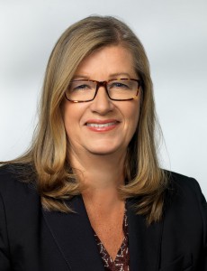
Prof. Katrine Riklund from Umeå, Sweden, will speak about the growing significance of hybrid imaging in today’s honorary lecture.

During her lecture, Prof. Riklund will share examples of what hybrid imaging can do and what is going to come next. A major trend will be making use of the entire hybrid imaging examination for diagnostic protocols, also for the CT part, she believes. “This changes workflow and gives us more information. PET and CT or MR are not competing techniques, they are complementary,” she said.

Tracer development is key for PET but digital detectors will also represent a major step forward. Currently, the need for a cyclotron for production of radionuclides hinders substantial distribution of scanners outside large centres. “To make hybrid imaging really take off, we need other forms of tracer production. I would like to see the tracer production work like a coffee machine – with buttons to select tracer and radionuclide,” she said.

In her day-to-day work, Prof. Riklund is involved in various research projects, such as COBRA, a prospective multimodal imaging study of dopamine, brain structure and function, and cognition; (PEARL-PD), 18F-FE-PE2I PET/CT, a study of dopamine transporters in early Parkinson’s disease, RECTOPET (REctal Cancer Trial On PET-MR/CT); and Prostate Cancer – PSMA and Acetate in PET/CT and PET/MR. These projects reflect her three major interests in the field; movement disorders and cognition in central nervous system, and prostate and colorectal cancer.
Read more…

Hybrid imaging contents to heighten delegates’ interest at ECR 2016

ECR2015_ECRToday_Blog

ECR Today spoke with ECR 2016 Congress President, Prof. Katrine Åhlström Riklund, deputy head of the department of radiation sciences and director of the medical school at Umeå University, Sweden, to find out a little bit about next year’s annual meeting.

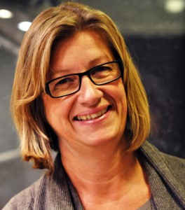
ECR 2016 Congress President, Prof. Katrine Åhlström Riklund, from Umeå, Sweden.

ECR 2016 Congress President, Prof. Katrine Åhlström Riklund, from Umeå, Sweden.

ECR Today: What will be the highlights of ECR 2016?
Katrine Åhlström Riklund: It is hard to tell what the specific highlights will be more than one year ahead, due to the rapid development of imaging. The highlights will be the entire congress through its well-developed programme, which covers the whole range of education from student level to advanced subspecialists. I should say the added content of hybrid imaging in several sessions would make the programme even more attractive. Besides the educational and scientific programme, the grand opening ceremony and social activities will also be memorable events.

ECRT: Will there be any new additions to the programme?
KAR: As always, there will be innovations at the ECR. The content of hybrid imaging will be spread across several sessions and not in one single session. The new session formats introduced at ECR 2015, with the European Excellence in Education (E3) programme – divided into five levels (the Rising Stars programme, European Diploma Prep Sessions and Beauty of Basic Knowledge programme, ECR Academies and ECR Master Classes) will be continued. These levels cover the entire span from undergraduate medical education to subspecialised continuing professional development. Getting involved in the sessions is important for retaining knowledge.

Read more…

Experts explain how to avoid pitfalls in FDG PET/CT imaging

ECR2014_ECRToday_Blog_Final

Watch this session on ECR Live: Thursday, March 6, 16:00–17:30, Room I/K
Tweet #ECR2014IK #SF3

The demand for PET/CT studies is increasing and so is the need for radiologists to improve their knowledge of this important modality. One of the many areas that require their attention is the occurrence of pitfalls related to the uptake of Fludeoxyglucose (18F), commonly called FDG, the most commonly used tracer in PET/CT imaging. A dedicated Special Focus session at the ECR will offer attendees useful clues on how to avoid these pitfalls and correctly interpret images.

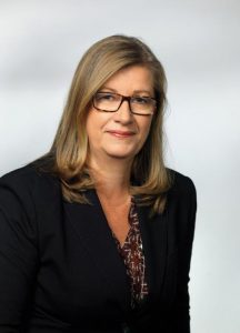
Katrine Åhlström Riklund is director of the medical school and deputy head of the department of radiation sciences at Umeå University, Sweden. She is 2nd vice-chairperson of the ESR’s Congress Committee.

Katrine Åhlström Riklund is director of the medical school and deputy head of the department of radiation sciences at Umeå University, Sweden. She is 2nd vice-chairperson of the ESR’s Congress Committee.

FDG uptake by tissue is also a marker for glucose uptake, which is closely correlated with certain types of tissue metabolism. This means that FDG can show not only disease-related changes but also normal, healthy metabolic changes in the body. “Not everything that shines is pathological. To know the difference, you have to train and learn what is really a disease and what is the physiological distribution of this tracer,” said Professor Katrine Åhlström Riklund, a radiologist specialised in nuclear medicine at Umeå, Sweden, who will moderate the session.
To help radiologists, speakers will share advice regarding FDG uptake in oncology, neurology and cardiology.

Most FDG PET/CT studies are currently being carried out to help stage cancer, and plan and follow-up therapy. The combination of FDG and PET/CT imaging is particularly useful in several different malignancies. Because a tumour cell divides rapidly and has a high rate of metabolism, FDG uptake usually corresponds to disease. Once physicians know the extent of the disease, they can make a more accurate diagnosis and treatment plan, especially in targeted therapies.

Read more…

06
Mar 2014
POSTED BY
POSTED IN ,
DISCUSSION 0 Comments