ECR 2018 Cases of the Day Winners

The winners of the ECR 2018 Cases of the Day Quiz are as follows:

Mehmet Serindere; Hatay/TR
Sungeun Park; Seoul/KR
Seungchul Han; Seoul/KR
Sewoo Kim; Seoul/KR
S. A. Sohaib; Sutton/UK
Rafal Darecki; Koscierzyna/PL

To view the cases please click here.

Congratulations to all winners!

26
Mar 2018
POSTED BY
POSTED IN ,
DISCUSSION 0 Comments

The best submissions for the “Normal variant or disease” interlude at ECR 2018

Dear Friends,

Over the last couple of years, one of the last sessions at the ECR has always covered 20 interesting cases from various subspecialties, which the audience is asked to solve in an interactive way to broaden and update their knowledge.

In between, the very best submissions from the global radiological community have been presented in an interlude lecture. The best submission has always been awarded with a prize and a certificate.

Due to time limits, not all submitted cases can actually be shown onsite, but the session’s rising popularity has resulted in increasing numbers of submissions of excellent quality and didactic value. This is why we would like to give our submitters the opportunity to reach a broader audience by posting the best cases here on the ESR Blog.

This year’s topic was Normal variant or disease to avoid misdiagnosis, overdiagnosis, unnecessary and costly work-up, as well as patient’s concerns.

When we think back of our time in medical school and remember our studies of embryology, we have probably all thought at some stage, how astonishing it is that a viable fetus develops and a healthy newborn is delivered. Embryological development is so complex that an awful lot can go wrong at any stage. Fortunately, real malformations are rare. In our professional life as radiologists, though, we are not infrequently dealing with normal variants, when development is arrested at a certain stage or turns the wrong way round. Even first year residents are familiar with right sided aortic arch, azygos lobe and accessory ossification centres. Over time, one encounters a whole array of normal variants and learns to interprete them as such. Of course, one can only identify, what one has learnt to see and there is a large spectrum of slight developmental abnormalities, which should not be misdiagnosed as abnormality in need of further imaging, invasive diagnostic procedures or treatment. On the other hand, some developmental disorders have clinical significance and you could be the first to advise the patient.

Below, we are pleased to present the best submissions for the “Normal variant or disease” interlude at ECR 2018:

Incoming ESR President Lorenzo Derchi looks forward to celebrating an ECR milestone in 2019


By Julia Patuzzi

As has been our tradition for a few years now on the final day of our congress, we already look ahead to next year’s ECR. We therefore spoke with Prof. Lorenzo E. Derchi from Genoa, Italy, who, as incoming ESR President, is in charge of ECR 2019. He told us about his plans and ideas for the next European Congress of Radiology.

ECR Today: Professor Derchi, next year’s European Congress of Radiology will be the 25th to be held in Vienna. Are there any specific celebrations planned that you can already share with us?

Prof. Lorenzo E. Derchi

As incoming ESR president, Prof. Lorenzo E. Derchi, Head of the Department of Radiology at the University of Genoa, will preside over ECR 2019.

Lorenzo E. Derchi: We are still thinking about them. An anniversary like this is something more than an opportunity for a big celebration. It is the right moment to re-think our history and what we have reached over the years. To always have the congress in the same city since 1991, at first every other year and then, after 1999, annually, has been a radical change. The new formula proved successful and ECR has become the European meeting to come to in order to learn the latest clinical and technical advances in our discipline as well as to present the results of the previous year’s research.

In my experience, however, it has become much more. From 1991 up until today all congress chairpersons have been part of the same team in a relay race, each handing off the baton to the next one. This continuity can be felt in the special atmosphere at each meeting, and has allowed the congress to become the annual appointment to meet old friends and make new ones; to discuss any radiological topic and then to start working together on it; and finally a place to meet periodically to share problems, advancements and results of long-term projects.

ECRT: Were you here for the first ECR in Vienna in 1991? If not, when did you attend your first ECR? What do you recall from that visit?

Read more…

Trojanowska to deliver honorary lecture on neglected form of cancer

Polish radiologist Dr. Agnieszka Trojanowska will shed light on human papilloma virus (HPV)-induced squamous cell cancer of the head and neck, a common yet long neglected type of cancer. In an interview with ECR Today, she also explained the origins of her passion for head and neck imaging, and how radiologists will increasingly benefit from computer science.

Dr. Agnieszka Trojanowska, assistant professor in the department of radiology and nuclear medicine at Lublin University Hospital, is an internationally recognised specialist in head and neck imaging. Her special interest for oncology and her natural curiosity led her to agree to deliver the Josef Lissner Honorary Lecture on HPV-induced squamous cell cancer of the head and neck (HNSCC) at ECR 2018.

Agnieszka Trojanowska from Lublin, Poland, will speak on HPV-induced squamous cell cancer of the head and neck in her Honorary Lecture today.

“HNSCC behaves differently than more well-known forms of SCC, and it is becoming more and more prevalent. It affects young, well-educated people with high socioeconomic status, and is a quickly rising sexually transmitted entity with peculiar clinical and molecular characteristics. In particular the rise of HPV-induced cancer has been observed in the United States since the beginning of 21st century,” she said.

It was important for her to talk about this long overlooked topic, for which much remains to be done. “It has been under-estimated for many years in many countries. Nowadays, we can speak about an HPV epidemic, leading to a significant rise of oropharyngeal cancer incidences worldwide. These cancers are quite unique and, in my opinion, knowledge of their physiology and treatment options is essential,” she said.

New research shows that, compared with environmental-related head and neck squamous cell carcinomas, patients with HPV-related malignancies display a better response to treatment and a lower risk of death and progression, Dr. Trojanowska pointed out. It is also noteworthy that, according to the 2017 TNM classification, HPV+ cancers are evaluated with a separate grading scale, she added.

Read more…

The past, the present and the future – a foray through Portuguese radiology


by Katharina Miedzinska

Saturday’s ‘ESR meets Portugal’ session is a successful combination of science, culture and entertainment, offering the opportunity to discover how Portugal has contributed to the practice of radiology over the years and foresee the future of Portuguese radiology.

In an interview with ECR Today, Dr. Filipe Caseiro Alves, professor at the University Clinic of Radiology, Coimbra University Hospitals, Portugal, outlined some central themes and highlights of Saturday’s highly anticipated session.

One of the first reports and image of cerebral angiography. From, Moniz E, de Carvalho L, Lima A. La radioartériographie et la topo-graphie cranioencephalique. J Radiol Electrol Med Nucl 1928;12:72. (Provided by Prof. Filipe Caseiro Alves)

At the beginning, Caseiro Alves plans to go on a short time travel through the history of Portuguese radiology. “Portugal’s greatest contributions to radiology are closely interwoven with the Portuguese School of Angiography, which began with the work of Egas Moniz, who was the first to perform a cerebral arteriography in 1927,” he explained.

Since then, Portugal has yielded many outstanding pioneers in the field of vascular radiology, among them Reynaldo dos Santos, a professor of surgery in Lisbon, who, among others, is known for the invention of aortography in 1929. Others include Lopo de Carvalho, who successfully introduced pulmonary angiography in 1931, Álvaro Rodrigues, Sousa Pereira and Roberto de Carvalho, who are known for their innovative work on lymphography (1933), Reynaldo’s son, João Cid dos Santos, who successfully introduced direct phlebography of the limbs in 1938, and Ayres de Sousa, whose name will always be closely associated with microangiography. “All these pioneer works were at the forefront of one of the most important achievements in healthcare and proved instrumental in shaping today’s medical practice,” Caseiro Alves said. Read more…

ECR continues to think outside the box and adds the Cube

For young physicians, interventional radiology (IR) may seem as enigmatic as a Rubik’s cube. But the cube can be solved with the right algorithm, according to Dr. Maximilian de Bucourt, head of angiography at Charité Campus Benjamin Franklin in Berlin.

“What we do in IR is exactly like a Rubik’s cube: you follow the algorithm and solve the problem. In IR, if you do the procedure steps over and over again and use the rules, most of the time you can get the solution for the patient,” he said.

Dr. Maximilian de Bucourt is head of angiography at Charité Campus Benjamin Franklin in Berlin, and one of the specialists behind the Cube.

Together with Prof. Christian Loewe from Vienna, de Bucourt imagined ‘the Cube’, a workshop aimed at introducing young physicians to the tools and techniques used in IR. With its focus on hands-on activities, including simulated procedures and interactive demonstrations, the Cube is fulfilling its goal of acquainting residents with this unique sub-specialty.

“The Cube is for young people who are thinking of becoming IR specialists, but find it too long before they can deploy a stent or manoeuver a catheter inside an artery. After medical school, basic radiology training and dedicated IR training with a teacher willing to let them perform major interventions, they still need to learn how to manage complications. The Cube is all about expediting this process, by enabling students and residents to get their hands on the products earlier,” he explained. Read more…

Riklund to unveil the next big thing in hybrid imaging

The Swedish radiologist Prof. Katrine Riklund, the current Chair of the ESR Board of Directors, has dedicated her career to the development of hybrid imaging. During todays’ Marie Curie Honorary Lecture (Room A, 1:00 p.m.) she will look back at the achievements made in this emerging field and look forward to future advances.

Riklund, who is a professor, consultant in diagnostic radiology and pro-vice-chancellor of Umeå University, is one of Sweden’s leading radiologists. She is also one of the first researchers to have recognised the potential of combining PET with CT and MRI, and has worked to advance the field ever since its emergence at the beginning of the 21st century.

“The combination of structural and functional/molecular imaging is fascinating. The entire field is new and extremely interesting, and it’s the closest to my heart when it comes to imaging,” she said.

Prof. Katrine Riklund from Umeå, Sweden, will speak about the growing significance of hybrid imaging in today’s honorary lecture.

During her lecture, Prof. Riklund will share examples of what hybrid imaging can do and what is going to come next. A major trend will be making use of the entire hybrid imaging examination for diagnostic protocols, also for the CT part, she believes. “This changes workflow and gives us more information. PET and CT or MR are not competing techniques, they are complementary,” she said.

Tracer development is key for PET but digital detectors will also represent a major step forward. Currently, the need for a cyclotron for production of radionuclides hinders substantial distribution of scanners outside large centres. “To make hybrid imaging really take off, we need other forms of tracer production. I would like to see the tracer production work like a coffee machine – with buttons to select tracer and radionuclide,” she said.

In her day-to-day work, Prof. Riklund is involved in various research projects, such as COBRA, a prospective multimodal imaging study of dopamine, brain structure and function, and cognition; (PEARL-PD), 18F-FE-PE2I PET/CT, a study of dopamine transporters in early Parkinson’s disease, RECTOPET (REctal Cancer Trial On PET-MR/CT); and Prostate Cancer – PSMA and Acetate in PET/CT and PET/MR. These projects reflect her three major interests in the field; movement disorders and cognition in central nervous system, and prostate and colorectal cancer.

Read more…

Swiss prepare to unveil connection between chocolate and radiology

by Viviene Raper

How to create hand-made chocolate masterpieces isn’t your usual radiology talk but, in tomorrow’s session, radiologists with a sweet tooth can learn all about truffle making and other confectionery arts from a leading Swiss chocolatier – interspersed with more conventional talks about radiology in Switzerland.

Andreas Trumpler, managing director marketing and group CMO at Läderach chocolatier suisse, will be giving two short presentations about how Swiss chocolate is transformed from cacao bean to chocolate, and then into a hand-made masterpiece. The company employs more than 800 people and since 1962, it has established a reputation for chocolate and confectionery specialties. Läderach’s products are sold throughout Switzerland and Germany, as well as by partners in the Middle East and Asia, and it supplies the top end of the gastronomy and hotel industry with semi-finished and finished products.

Fresh Chocolate with almonds from Läderach chocolatier suisse: the original view.

The connection between Swiss chocolate and Swiss radiology is one of quality, according to Prof. Dr. Dominik Weishaupt, who has been President of the Swiss Society of Radiology (SGR-SSR) since 2016 and is chairing tomorrow’s session.

“Over the 100 years of the Society’s history, we have undertaken several initiatives to improve the quality of Swiss radiology. And chocolate is considered as one of the highest quality products manufactured in Switzerland,” he noted.

Weishaupt, who is chief physician in the Institute of Radiology and Nuclear Medicine at City Hospital Zurich, explained that Swiss chocolate needs precision manufacturing, and Swiss radiology also prides itself on its attention to detail. He said that the SGR-SSR was the first medical society to introduce board exams, as well as clinical audits for radiology departments in the private and public sector, which they developed in close collaboration with the Ministry of Health. Read more…

The future is now, Marc Dewey says

The radiological community must understand the potential of value-based radiology and its related challenges, the German radiologist Marc Dewey will argue during the Wilhelm Conrad Röntgen honorary lecture today at ECR 2018.

Marc Dewey is Heisenberg Professor of Radiology of the German Research Foundation and Vice Chair of the Department of Radiology at Charité – Universitätsmedizin Berlin. His main research interest is in cardiovascular imaging but he is interested in many other aspects of clinical practice including value-based imaging, something he will detail in his lecture at the ECR.

Prof. Marc Dewey from Berlin will deliver today’s Honorary Lecture on value-based imaging at 12:15 in Room A.

“Participants will get to know three things. First, why the time for value-based radiology is now. Second, they will appreciate the potential of value-based radiology in the clinical care process. And third, understand the challenges in implementing value-based radiology,” Dewey promised.

Combining human image analysis and artificial intelligence has great potential for creating value for patients at lower costs, he believes. “This is value-based radiology and the time to get involved is now, as this is not merely a new technical toy of radiologists but will be accompanying the entire diagnostic and treatment pathway in all clinical service lines for the benefit of our patients,” he said.

Dewey is the coordinator of the DISCHARGE trial, an EU-funded multicentre project with more than 30 partners across Europe that will determine whether cardiac CT may replace invasive coronary angiography in certain patients. “The DISCHARGE project is a unique and truly impressive effort of several hundred individuals working at 31 sites in 18 European countries. External advisory board members were often thrilled when first witnessing the team spirit at our annual meetings,” said Dewey, who will soon present results of the on-going clinical trial. He said coordinating a large project on imaging in Europe gave him “the ability to better understand the culture, concerns, and ideas in different regions of Europe.”

Read more…

ECR 2018 celebrates the diversity of radiology

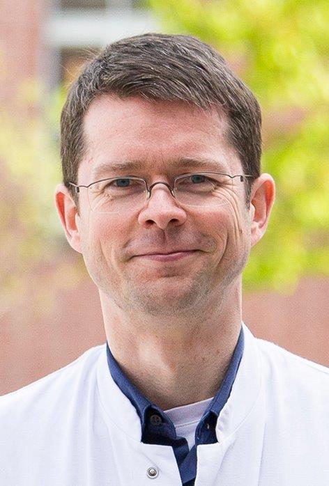
by Prof. Bernd Hamm, ESR President

It is my great pleasure to welcome you to ECR 2018!

The European Congress of Radiology has attendees from all over the world – from over 140 countries and many diverse cultures. It is a multi-professional meeting where international experts can shake hands with students, medical residents exchange ideas with physicists, and radiographers share their perspectives with industry representatives.

I chose ‘Diverse & United’ as our congress motto this year, as radiology is such a diverse specialty, covering a huge range of medical and scientific topics: from ever more refined diagnostic options to image-guided minimally invasive treatment options. Alongside our diversity, as radiologists and radiographers we should also stay united, which is in the interest of our specialty and our patients. This is what our congress is: something to offer for everyone, regardless of profession, cultural background or specialisation.

Bernd Hamm, ESR President

ESR President Bernd Hamm is professor of radiology and chairman of all three merged departments of radiology at the Charité, Humboldt-Universität zu Berlin and Freie Universität. He is also clinical director of the Charité Center, which includes radiology, neuroradiology, nuclear medicine and medical physics.

Being Congress President in 2015 gave me the unique opportunity for re-evaluation and to implement new things that I feel strongly about as well as fine-tune features that already existed. It was generally important to me to introduce new ideas in order for those of you who’ve even been coming to Vienna for decades, just like me, to get the chance to experience multiple innovations.

MyT3 is a new session format, adapting the ECR even more to these fast-moving times. 240 daring colleagues will present their scientific thesis in just three minutes! As if this wasn’t dramatic enough, we decided to hold these speedy sessions on the Sky High Stage which overlooks the city of Vienna, as only the sky is the limit for this new generation of radiology professionals.

Additionally, we created another new session format, ‘Coffee & Talk’, which is highly interactive with much more time for discussion than usual and in a relaxed atmosphere, with the possibility to also enjoy a coffee or other hot beverage. The interesting lectures on offer in these sessions call for an exciting exchange, bringing together different statements and opinions as well as Viennese coffee culture.

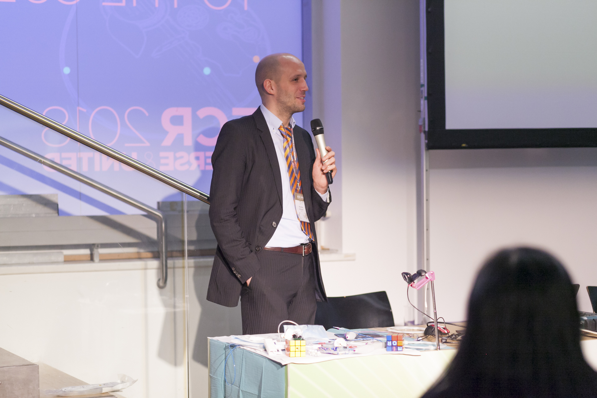
For the first time, the CUBE will open its doors to you: a theme park for interventional radiology (IR), designed for residents who haven’t specialised yet. Challenges, quizzes, training and much more will be focused on IR in emergencies plus other everyday topics, including the aorta, oncology, peripherals, and stroke. Without wanting to give away too much, I recommend paying the Cube a visit during lunchtime for ‘the main event’: the daily highlight involving experts in the arena, less challenging as well as more challenging interventions and much more. Come and be part of it!

Read more…