CEUS ready for clinical paediatric use argues UK expert

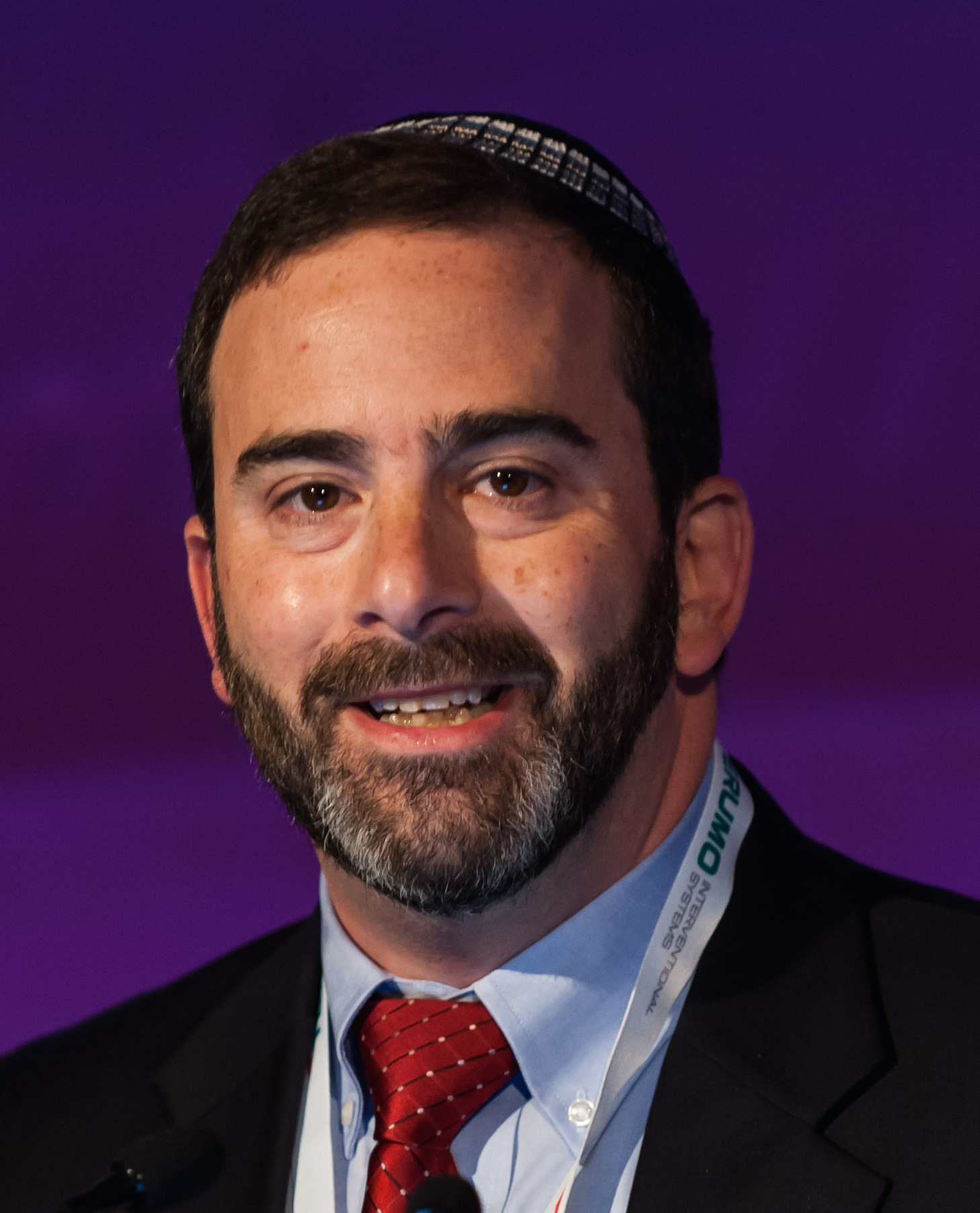
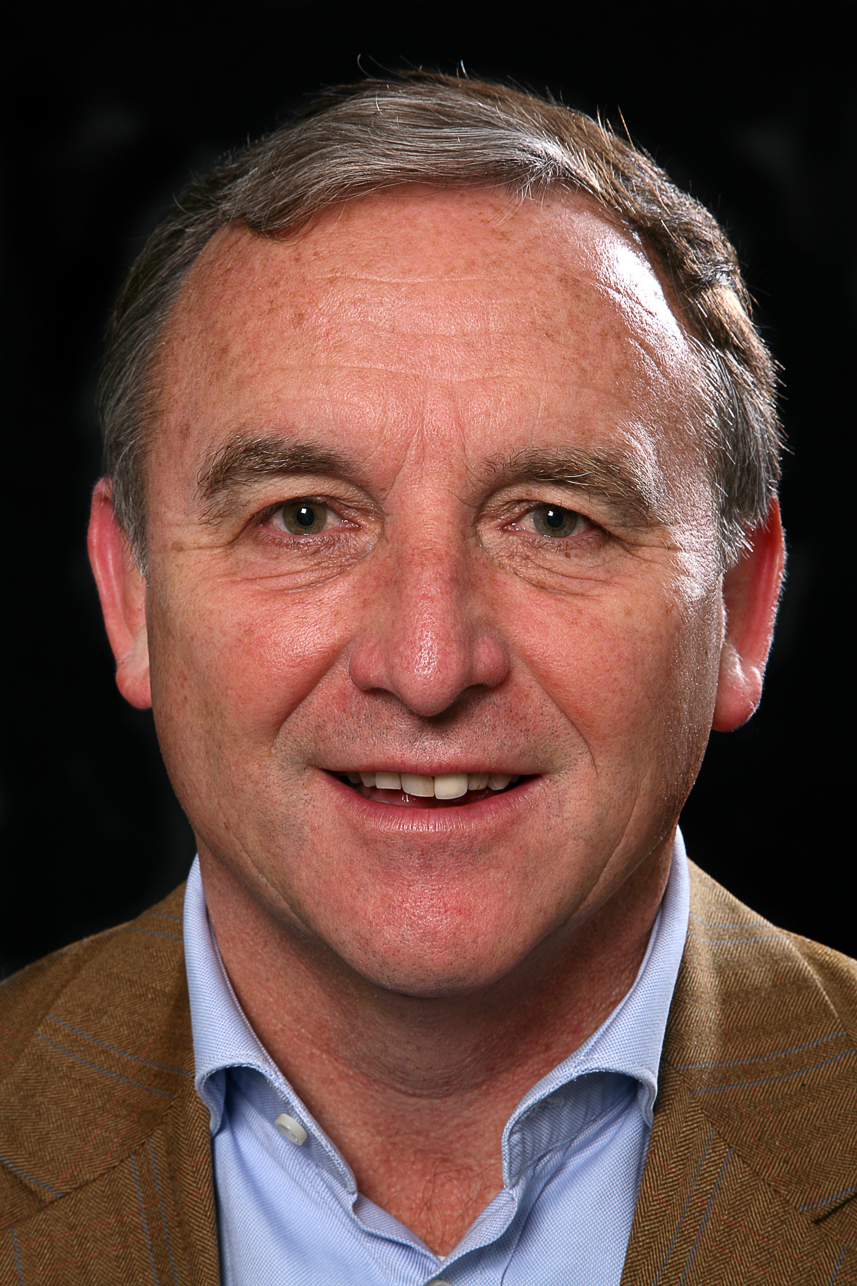
Professor Paul Sidhu is Professor of Imaging Sciences at King’s College London and consultant radiologist at King’s College Hospital, where he helped pioneer contrast-enhanced ultrasound (CEUS), a technique that can potentially be used to image children just as well as adults, in reduced time, and without the downsides of radiation and toxicity. Professor Sidhu will make the case for CEUS use in paediatric radiology today during the Luigi Oliva Honorary Lecture.

There is no question that ultrasound is the most child-friendly imaging technique. The modality has countless benefits for young patients: no ionising radiation, no sedation or anaesthesia, and ease of use, to name a few. “Ultrasound is the most suitable modality for imaging children, as they don’t have to stay still. Parents can be present during the examination to support their child. The radiologist can stop and start the examination without losing information, plus children have low body fat and are better suited to ultrasound,” Sidhu said.

Professor Paul Sidhu from King’s College London will argue for CEUS use in paediatric radiology in today’s Luigi Oliva Honorary Lecture.

Adding a contrast agent to ultrasound has been shown to improve diagnosis tremendously in applications where vascularity or haemo-dynamics must be assessed in real time. With CEUS, in the case of focal liver lesions, which are often difficult to characterise in adults and children on B-mode ultrasound alone, essential information from the arterial and portal venous phases aids diagnosis in minutes without needing CT or MR imaging. This alone should be a decisive argument for widespread CEUS use in the paediatric population, Sidhu explained.

“The combination of contrast with ultrasound allows for a more detailed assessment of the pattern of vascularity and dynamics in a continuous fashion for three to five minutes in real time, and it can be repeated as many times as necessary. The child does not need to keep still, and all the same focal liver characteristics seen in the adult are seen in the child. The ability to come to a clear diagnosis, with a single imaging examination is a great relief for the parents, often present with the child during the examination, rather than remote as with CT or MR imaging,” he said.

With CEUS, radiologists can obtain a lot of information in the first five minutes following the injection, the time window that the contrast agent lasts in the blood pool. This is more than enough to carry out the examination and make a conclusion. The technique can be used in every possible scenario from trauma to disease imaging, with great results. “Imaging with ultrasound in children is the most useful and informative procedure,” Sidhu concluded. Read more…

Systemic effects in interventional oncology: Holy Grail or Pandora’s Box?

Nahum Goldberg is full professor of radiology at Hadassah Hebrew University, Jerusalem, Israel and visiting professor of radiology at Harvard Medical School, Boston, MA. He is a worldwide expert in image-guided tumour therapy, a field he has helped pioneer and continues to advance. He will share some of the latest results on the effects of these therapies, both positive and negative, today during the Josef Lissner Honorary Lecture.

Image-guided therapy and transcatheter intervention work best on small tumours in the liver, kidney and several other organs, evidence over the past two decades has showed. That was the birth of interventional oncology and it held exciting promise for the (radical) improvement of cancer treatment. Based on that knowledge and promise, many researchers have worked towards combining these procedures with more conventional cancer therapies like chemotherapy and radiation to increase the size and range of the population that can be treated.

Prof. S. Nahum Goldberg from Jerusalem will share some of the latest results on the effects of image-guided tumour therapy, during the Honorary Lecture today at 12:15.

One of the underlined premises of interventional therapies is that using imaging to guide a procedure is less invasive than using other methods like surgery. Despite this and other advantages that have led to ablation systems and techniques now being used clinically more than 100,000 times annually, things have not always gone as planned, according to Goldberg, who heads the interventional oncology unit at Hadassah Medical Centre.

“Specifically, for much of the last two decades, we have argued that other benefits of these interventional oncologic therapies, including percutaneous tumour ablation and chemoembolisation, were focal and local, based upon our conviction that we were only affecting the tumour we were treating and not the entire patient system. It turns out, based on early case reports and more solid evidence produced over the last five years or so, that this premise is not always true,” said Goldberg.

“Most of the damage occurs where intended, but all kinds of pathways in the system are also activated after an intervention. Just like a sunburn that affects a local piece of skin can be accompanied by a headache or fever, a systemic reaction can follow an interventional oncologic procedure in some patients and under certain circumstances. Depending on the type of tumour and its location, a series of both positive and negative systemic effects can be unleashed,” he added.

These effects are rather variable and are currently hard to predict, but one thing is certain: there is a good side and a bad side – the Holy Grail and Pandora’s Box, as Goldberg put it. Read more…

A new beginning in cancer imaging has just begun, says Beets-Tan

Professor Regina Beets-Tan is chair of the department of radiology at The Netherlands Cancer Institute in Amsterdam, full professor of radiology at the University of Maastricht and adjunct professor of abdominal and oncological radiology at the University of Southern Denmark. She will present the Wilhelm Conrad Röntgen Honorary Lecture, titled ‘Oncologic imaging: a new beginning has just begun’ at ECR 2019.

She shared a few thoughts with us on the future of her specialty in an interview ahead of the congress.

Prof. Regina Beets-Tan from Amsterdam will talk about future aspects of oncologic imaging in today’s honorary lecture.

ECR Today: You have chosen quite an iconic and broad topic for this honorary lecture. What points will you cover exactly?

Regina Beets-Tan: The audience will get a glimpse of the future of cancer care and the role of imaging. The world of cancer medicine is changing rapidly. Major steps forward have been taken. Advanced imaging and computing technology, screening programmes; these all will result in the early detection of more tumours. Minimally invasive treatment, including interventional therapy, will have an increasingly important role. Targeted therapy, which specifically hits the cancer genes, and immunotherapy, which uses the patient’s own immune system to kill cancer cells, will result in prolonged survival of patients who are in the final stage of metastatic disease. It will be ‘precision medicine’; we do not want to give the wrong treatment to the wrong patient. As advocated by Prof René Bernards, a respected leader in cancer research at the Netherlands Cancer Institute: ‘Within 15 years, cancer will become a chronic disease’. And I believe this is true. This transformation will change the way we will practice oncologic imaging. This will require us to recreate our discipline. With this lecture, I would like to take my young colleagues on a 20-minute journey towards their future. Read more…

Trojanowska to deliver honorary lecture on neglected form of cancer

Polish radiologist Dr. Agnieszka Trojanowska will shed light on human papilloma virus (HPV)-induced squamous cell cancer of the head and neck, a common yet long neglected type of cancer. In an interview with ECR Today, she also explained the origins of her passion for head and neck imaging, and how radiologists will increasingly benefit from computer science.

Dr. Agnieszka Trojanowska, assistant professor in the department of radiology and nuclear medicine at Lublin University Hospital, is an internationally recognised specialist in head and neck imaging. Her special interest for oncology and her natural curiosity led her to agree to deliver the Josef Lissner Honorary Lecture on HPV-induced squamous cell cancer of the head and neck (HNSCC) at ECR 2018.

Agnieszka Trojanowska from Lublin, Poland, will speak on HPV-induced squamous cell cancer of the head and neck in her Honorary Lecture today.

“HNSCC behaves differently than more well-known forms of SCC, and it is becoming more and more prevalent. It affects young, well-educated people with high socioeconomic status, and is a quickly rising sexually transmitted entity with peculiar clinical and molecular characteristics. In particular the rise of HPV-induced cancer has been observed in the United States since the beginning of 21st century,” she said.

It was important for her to talk about this long overlooked topic, for which much remains to be done. “It has been under-estimated for many years in many countries. Nowadays, we can speak about an HPV epidemic, leading to a significant rise of oropharyngeal cancer incidences worldwide. These cancers are quite unique and, in my opinion, knowledge of their physiology and treatment options is essential,” she said.

New research shows that, compared with environmental-related head and neck squamous cell carcinomas, patients with HPV-related malignancies display a better response to treatment and a lower risk of death and progression, Dr. Trojanowska pointed out. It is also noteworthy that, according to the 2017 TNM classification, HPV+ cancers are evaluated with a separate grading scale, she added.

Read more…

Riklund to unveil the next big thing in hybrid imaging

The Swedish radiologist Prof. Katrine Riklund, the current Chair of the ESR Board of Directors, has dedicated her career to the development of hybrid imaging. During todays’ Marie Curie Honorary Lecture (Room A, 1:00 p.m.) she will look back at the achievements made in this emerging field and look forward to future advances.

Riklund, who is a professor, consultant in diagnostic radiology and pro-vice-chancellor of Umeå University, is one of Sweden’s leading radiologists. She is also one of the first researchers to have recognised the potential of combining PET with CT and MRI, and has worked to advance the field ever since its emergence at the beginning of the 21st century.

“The combination of structural and functional/molecular imaging is fascinating. The entire field is new and extremely interesting, and it’s the closest to my heart when it comes to imaging,” she said.

Prof. Katrine Riklund from Umeå, Sweden, will speak about the growing significance of hybrid imaging in today’s honorary lecture.

During her lecture, Prof. Riklund will share examples of what hybrid imaging can do and what is going to come next. A major trend will be making use of the entire hybrid imaging examination for diagnostic protocols, also for the CT part, she believes. “This changes workflow and gives us more information. PET and CT or MR are not competing techniques, they are complementary,” she said.

Tracer development is key for PET but digital detectors will also represent a major step forward. Currently, the need for a cyclotron for production of radionuclides hinders substantial distribution of scanners outside large centres. “To make hybrid imaging really take off, we need other forms of tracer production. I would like to see the tracer production work like a coffee machine – with buttons to select tracer and radionuclide,” she said.

In her day-to-day work, Prof. Riklund is involved in various research projects, such as COBRA, a prospective multimodal imaging study of dopamine, brain structure and function, and cognition; (PEARL-PD), 18F-FE-PE2I PET/CT, a study of dopamine transporters in early Parkinson’s disease, RECTOPET (REctal Cancer Trial On PET-MR/CT); and Prostate Cancer – PSMA and Acetate in PET/CT and PET/MR. These projects reflect her three major interests in the field; movement disorders and cognition in central nervous system, and prostate and colorectal cancer.

Read more…

The future is now, Marc Dewey says

The radiological community must understand the potential of value-based radiology and its related challenges, the German radiologist Marc Dewey will argue during the Wilhelm Conrad Röntgen honorary lecture today at ECR 2018.

Marc Dewey is Heisenberg Professor of Radiology of the German Research Foundation and Vice Chair of the Department of Radiology at Charité – Universitätsmedizin Berlin. His main research interest is in cardiovascular imaging but he is interested in many other aspects of clinical practice including value-based imaging, something he will detail in his lecture at the ECR.

Prof. Marc Dewey from Berlin will deliver today’s Honorary Lecture on value-based imaging at 12:15 in Room A.

“Participants will get to know three things. First, why the time for value-based radiology is now. Second, they will appreciate the potential of value-based radiology in the clinical care process. And third, understand the challenges in implementing value-based radiology,” Dewey promised.

Combining human image analysis and artificial intelligence has great potential for creating value for patients at lower costs, he believes. “This is value-based radiology and the time to get involved is now, as this is not merely a new technical toy of radiologists but will be accompanying the entire diagnostic and treatment pathway in all clinical service lines for the benefit of our patients,” he said.

Dewey is the coordinator of the DISCHARGE trial, an EU-funded multicentre project with more than 30 partners across Europe that will determine whether cardiac CT may replace invasive coronary angiography in certain patients. “The DISCHARGE project is a unique and truly impressive effort of several hundred individuals working at 31 sites in 18 European countries. External advisory board members were often thrilled when first witnessing the team spirit at our annual meetings,” said Dewey, who will soon present results of the on-going clinical trial. He said coordinating a large project on imaging in Europe gave him “the ability to better understand the culture, concerns, and ideas in different regions of Europe.”

Read more…

Highly-respected Dutch researcher delivers honorary lecture on cardiac imaging

ECR2014_ECRToday_Blog_Final

In recognition of his significant research and work in the areas of MRI, CT and cardiovascular imaging, Professor Albert de Roos from Leiden, the Netherlands, was invited to deliver the Josef Lissner Honorary Lecture entitled, ‘Research in cardiac imaging: how I do it’.

Prof. Albert de Roos from Leiden, the Netherlands.

Prof. Albert de Roos from Leiden, the Netherlands.

Albert de Roos is professor of radiology at Leiden University Medical Center in the Netherlands. Originally from Ermelo, the Netherlands, Prof. de Roos received his medical degree (Cum Laude) from the University of Amsterdam in 1980 and then completed his residency in internal medicine at Zeeweg Hospital Velsen. He then went on to carry out a residency in radiology at St. Elisabeth Gasthuis Haarlem and University Hospital Leiden. In 1985, he was awarded a PhD for his thesis on ‘Biphasic Colon Examination’.

In 1988, Prof. de Roos travelled to the United States to become assistant professor of radiology at the University of Pennsylvania, Philadelphia. He returned to the Netherlands to become associate professor of radiology at Leiden University Medical Center, where he has held a number of posts, including director of magnetic resonance imaging and director of computed tomography. He also served as co-leader of the Interuniversity Cardiology Institute of the Netherlands’ ‘Cardiovascular MRI’ project.

Read more…