Radiology in the era of personalised medicine: is radiology personalised enough?

iStock_000006149013_Double_small

To select the right treatment at the right time for your patient is a common slogan used to describe personalised medicine (PM): a new vision of healthcare which is already becoming a reality today. Different “omics” approaches such as genomics, proteomics or metabolomics allow patients’ predispositions to certain diseases or treatment response to specific medication to be predicted.

What is the role of radiology and radiologists in PM?
In the recent white paper, published by the ESR Working Group on Personalised Medicine (Insights Imaging (2015) 6:141–155), the authors emphasise the key roles medical imaging plays in PM. The authors clearly specify all areas and give examples of the unique and personalised information provided with medical imaging technologies.

In a substantial number of diseases, the first step leading from clinical symptoms to a diagnosis relies on imaging. Imaging has played this role for decades: assessing the location and extent of a disease in individual patients and characterising structural abnormalities and physiological environment, thus providing crucial information for the choice of the right treatment.
Read more…

Comprehensive personalised imaging transforms cardiothoracic disease management

ECR2015_ECRToday_Blog

Watch this session on ECR Live: Sunday, March 8, 8:30–10:00, Room E1
Tweet #ECR2015E1 #NH17

Besides personalised imaging, a new paradigm is emerging in radiology that should re-shape clinical practice and benefit the patient immensely. Supported by new technologies that enable radiologists to image the body faster and better, radiologists are now trying to broaden their focus during examinations.

If there is a field where these advances make a tremendous difference, it is cardiothoracic imaging, an area where diseases are more often than not intertwined. Cardiovascular and chest radiologists will explain how the comprehensive personalised approach impacts their work and try to convince radiologists on both sides to take an interest in the other, in a New Horizons session on Sunday at the ECR.

For years, the trend was for radiologists to subspecialise as much as they could. Cardiovascular radiologists and chest radiologists would focus on their own area with little or no interest beyond that. But among these subspecialists, an increasing number are now changing their approach, as mounting evidence shows that diseases of the heart and chest are very often related, according to Dr. Christian Loewe, deputy head of the section of cardiovascular and interventional radiology at the Medical University of Vienna, Austria.

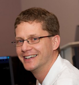
Dr. Christian Loewe is deputy head of the cardiovascular and interventional radiology section at the Medical University of Vienna, Austria.

Dr. Christian Loewe is deputy head of the cardiovascular and interventional radiology section at the Medical University of Vienna, Austria.

“In the past patients were investigated by either focusing on chest or cardiac diseases. This choice was mainly driven by their first clinical examination. However, there are a lot of situations and diseases where chest problems are caused by cardiac diseases and vice versa. There’s a huge interaction between heart and chest, and that’s why it’s interesting and important to look at this relationship in more detail today,” he said.

To prove his point, Loewe, a cardiovascular radiologist, will talk about acute and chronic chest pain during the session. Some of the most severe causes of chest pain are due to cardiovascular diseases, including myocardial infarction or acute aortic diseases. However, acute chest pain can also be caused by a number of pulmonary diseases, including pulmonary embolism, pneumonia and others. Therefore, radiologists must learn the different life-threatening disorders that cause chest pain, whether they are respiratory or cardiovascular.

Read more…

ESR initiatives to strengthen the visibility and role of imaging in personalised medicine

ECR2014_ECRToday_Blog_Final

An interview with Prof. Hans-Ulrich Kauczor, Heidelberg/DE, ESR Research Committee Chair

ECR Today: What are the Research Committee’s objectives?
Hans-Ulrich Kauczor: Our main tasks are to periodically survey the needs of the research community, as well as develop recommendations for radiologists to engage with different fields of research and innovation. We also work to assess the current status and develop recommendations on how to improve education in research and to provide strategic recommendations for the research field to the executive council. Last but not least, we aim to leverage cooperation between research disciplines and foster networking and liaising with scientific biomedical European societies.

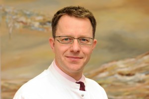
Hans-Ulrich Kauczor is professor and chairman of radiology at the University of Heidelberg and director of diagnostic and interventional radiology at University Hospital Heidelberg in Germany. He chairs the ESR’s Research Committee.

Hans-Ulrich Kauczor is professor and chairman of radiology at the University of Heidelberg and director of diagnostic and interventional radiology at University Hospital Heidelberg in Germany. He chairs the ESR’s Research Committee.

ECRT: Why is it important to have a dedicated ESR subcommittee for imaging biomarkers?
HUK: The Subcommittee on Imaging Biomarkers was established to address the issues concerning the future development of image-derived quantitative biomarkers, its assessment, validation and standardisation.

The development of imaging biomarkers has become an integral part of modern medicine with a huge potential to advance the development of personalised medicine. Different types of imaging biomarkers (anatomical, functional, and molecular) are used for the detection and treatment of major diseases including cancer, cardiovascular diseases, neurological and psychiatric diseases, musculoskeletal disorders, metabolic diseases, as well as inflammatory and autoimmunity based diseases. In contrast to other biomarkers, imaging biomarkers have the advantage of remaining non-invasive. They are also spatially and temporally resolved, non-destructive and repeatable over a long period, and have the potential for broad application. But before imaging biomarkers can be widely adopted, measures for standardisation and quality assurance must be implemented.

Read more…

ESR set to tackle personalised medicine and face economic realities in 2013

ECR2013_ECRToday_Monday_realities2013

In an interview with ECR Today, incoming ESR President, Professor Guy Frija explained how the ESR should address the challenges raised by the explosion of ‘omics’ data and the advent of personalised medicine, as well as how it should assess the impact of the financial crisis on the specialty.

ECR Today: What are your plans and ambitions regarding your presidency?
Guy Frija: The development of the ESR since its establishment in 2005 has been amazing: the ESR is now the biggest radiological society in the world, and its annual meeting, the European Congress of Radiology (ECR), is one of the best radiology congresses worldwide. All of this is the result of intense activity in several fields, such as education, research, professional challenges and publications, for the development of which the role of my predecessors was instrumental, along with the dedication of many of our colleagues. Therefore my first goal is to keep up this momentum and run the society appropriately, in accordance with its new statutes. However, life is challenging, and we will have to face many new issues in the coming year.

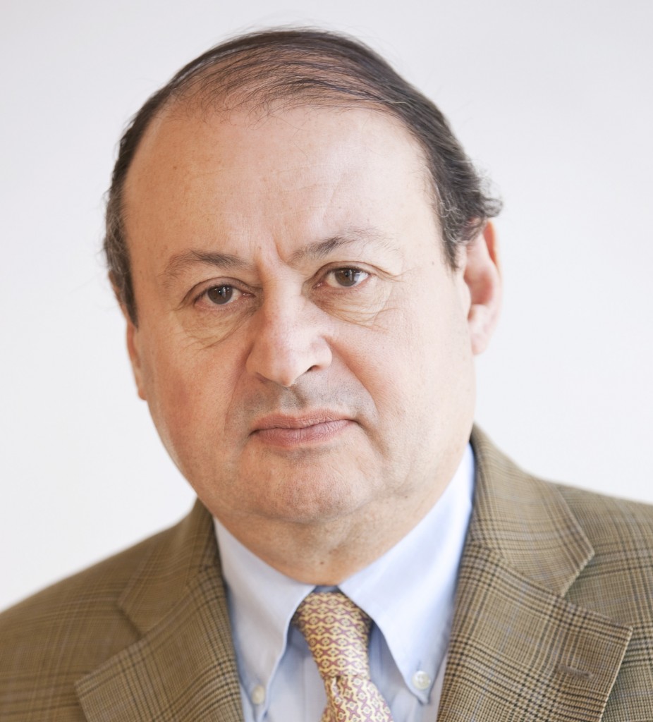
Professor Guy Frija is head of the imaging department at the Georges Pompidou European Hospital (Hôpital Européen Georges Pompidou, H.E.G.P.) in Paris.

Professor Guy Frija is head of the imaging department at the Georges Pompidou European Hospital (Hôpital Européen Georges Pompidou, H.E.G.P.) in Paris.

The explosion of ‘omics’ (such as metabolomics, proteomics, genomics) data is changing the face of medicine so considerably and rapidly that, if we don’t pay enough attention, our specialty could be threatened. Personalised medicine is not a just a dream, but an actual reality, and we have to adapt our specialty to this new paradigm. This year, DNA sequencing could become available for less than €750 (US$1,000), and in the UK, the Prime Minister David Cameron has launched a programme for the development of personalised medicine. The development of personalised imaging, which has already begun in oncology, should be taken further, and we have to appreciate its impact on education. I will ask two or three subspecialties to reconsider their training charters in light of these new developments. The data explosion related to these changes raises not only the problem of their management, but also of their interpretation in an integrated way: the imaging parameters should be included in this new data knowledge organisation, where knowledge modelling will become a major challenge. The establishment of imaging biobanks should be promoted along with adapted data privacy regulation.

Read more…

11
Mar 2013
POSTED BY
POSTED IN , ,
DISCUSSION 0 Comments