 Dear friends,
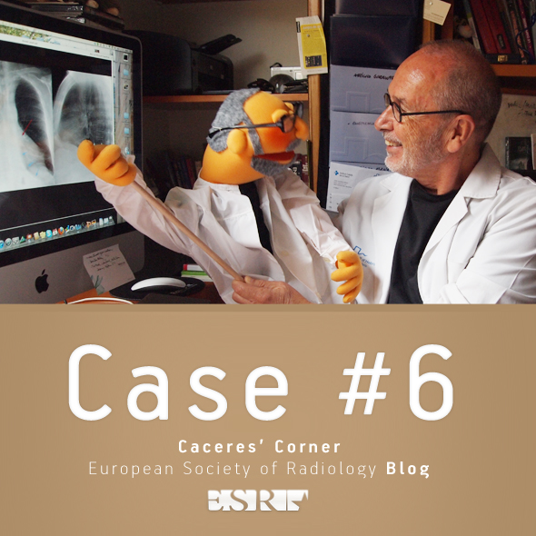
Welcome to Case #6. Let’s see what you make of this…
Clinical history: pre-operative chest radiograph for rhinoplasty in a 28-year-old male.
Diagnosis:
1. Middle lobe disease
2. Pleural effusion
3. Lung mass
4. None of the above
Read more…

Dear friends,
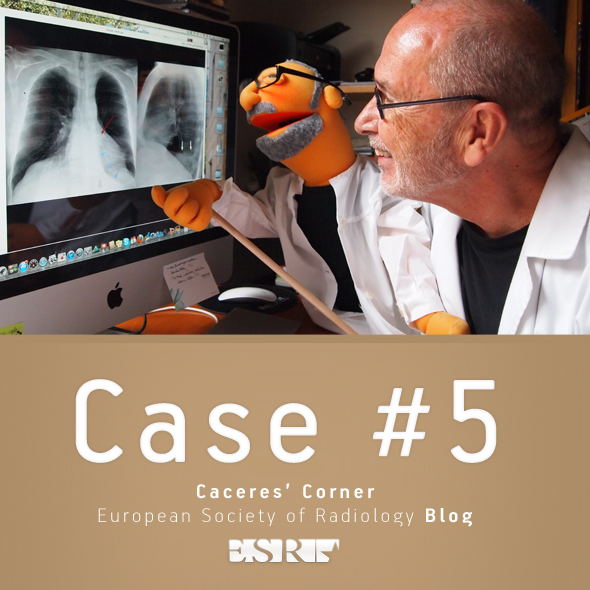
Welcome to case #5.
Case #5 is also from my wife. It is a 27-year-old female with a chronic infiltrate in the right upper lobe. My Wife asked us (ordered, according to Muppet) to help her with the case, because she doesn’t know what it is.
Diagnosis:
1. Tuberculosis
2. Aspiration pneumonia
3. Bronchioalveolar carcinoma
4. Lymphoma
I didn’t make the diagnosis, but the Muppet did! What is the Muppet’s diagnosis?
Read more…

Dear friends,
Welcome back to Caceres’ corner.
I’m already keen on your suggestions on Case #4.
Clinical history: Pre-operative chest radiograph for ophthalmologic surgery in a 57 y. o. male
Most likely diagnosis:
1. Carcinoma of the lung
2. Unilateral hyperlucent lung
3. Pulmonary embolism
4. Giant bulla
Read more…

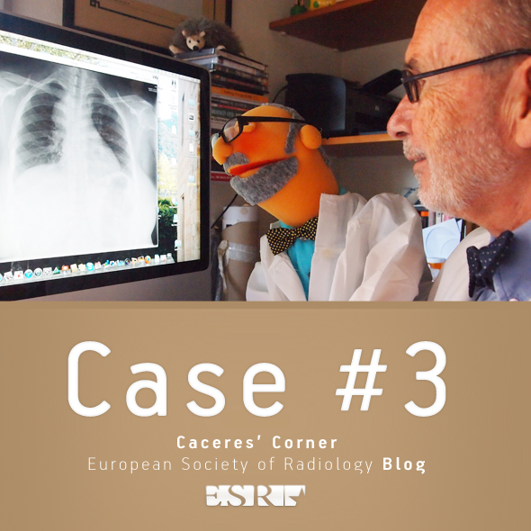
Welcome to case number three!
My wife, who works in a primary care centre, sent this case to me. The patient is a 76 year-old male, who was operated on three years ago for carcinoma of the larynx. The PA and lateral radiographs show two nodular lesions in the lower lung fields.
The obvious response is metastatic disease. But the Muppet, trying to impress both of us, suggested an alternative diagnosis.
Can you guess the Muppet’s diagnosis?
Read more…
 Clinical history: asymptomatic 75 year-old man, operated on ten years ago for carcinoma of the larynx. Current chest radiographs were obtained during the yearly routine control.
Most likely diagnosis:
1. Radiation pneumonitis
2. Tuberculosis
3. Carcinoma of the lung
4. Pulmonary hypertension Read more…

Meet Professor Jose Caceres and his Muppet
Dear friends, welcome to Caceres’ Corner. The objective of this post is to remember basic principles of chest imaging, with the emphasis on conventional radiography. Interpreting a chest radiograph is becoming a lost art and I would like to slow this tendency by reviewing the current approach to chest x-ray.
Nowadays, the initial question when facing a chest radiograph should be: “is there any abnormality present? And, if so, should we do any additional examination?” (CT in the great majority of cases).
With this approach in mind, let’s start with a sample case: 62 year old male with liver cirrhosis and upper gastrointestinal bleeding. No other symptoms. History of pulmonary tuberculosis 20 years ago.
Read more…


