Swiss prepare to unveil connection between chocolate and radiology

by Viviene Raper

How to create hand-made chocolate masterpieces isn’t your usual radiology talk but, in tomorrow’s session, radiologists with a sweet tooth can learn all about truffle making and other confectionery arts from a leading Swiss chocolatier – interspersed with more conventional talks about radiology in Switzerland.

Andreas Trumpler, managing director marketing and group CMO at Läderach chocolatier suisse, will be giving two short presentations about how Swiss chocolate is transformed from cacao bean to chocolate, and then into a hand-made masterpiece. The company employs more than 800 people and since 1962, it has established a reputation for chocolate and confectionery specialties. Läderach’s products are sold throughout Switzerland and Germany, as well as by partners in the Middle East and Asia, and it supplies the top end of the gastronomy and hotel industry with semi-finished and finished products.

Fresh Chocolate with almonds from Läderach chocolatier suisse: the original view.

The connection between Swiss chocolate and Swiss radiology is one of quality, according to Prof. Dr. Dominik Weishaupt, who has been President of the Swiss Society of Radiology (SGR-SSR) since 2016 and is chairing tomorrow’s session.

“Over the 100 years of the Society’s history, we have undertaken several initiatives to improve the quality of Swiss radiology. And chocolate is considered as one of the highest quality products manufactured in Switzerland,” he noted.

Weishaupt, who is chief physician in the Institute of Radiology and Nuclear Medicine at City Hospital Zurich, explained that Swiss chocolate needs precision manufacturing, and Swiss radiology also prides itself on its attention to detail. He said that the SGR-SSR was the first medical society to introduce board exams, as well as clinical audits for radiology departments in the private and public sector, which they developed in close collaboration with the Ministry of Health. Read more…

ECR 2013 Focus: Imaging of the Mind

ECR2013_Focus_7_ImagingMind

The brain remains undoubtedly one of the most mysterious organs of the human body. Magnetic resonance imaging has helped to unveil some of its secrets, and major advances have been made in understanding how the brain functions. Recent developments with resting fMRI (rfMRI) and diffusion MRI (dMRI) indicate that scientists are beginning to see beyond the brain: they have actually started to visualise the human mind. This new information is particularly relevant for understanding complex processes such as dementia, autism and depression. It is also proving increasingly central to the diagnosis of comas and chronic disorders of consciousness.

Leading researchers will discuss where the latest advances have led them and what the future will bring in a dedicated New Horizons Session during ECR 2013. FMRI has been used for over twenty years to visualise changes in brain activity by comparing a task versus a control task, and showing and quantifying how much brain activity is involved in the process. The recent addition of rfMRI enables researchers to track networks that are randomly active. A patient lying in a scanner with no particular task to perform will usually start thinking about the trivialities of the day and go from one thought to the other (“Did I close the door before I left? What am I doing here?” etc.). Neuroresearchers can track this mind mumbling with complex mathematics and extract information from what they call the default mode network.

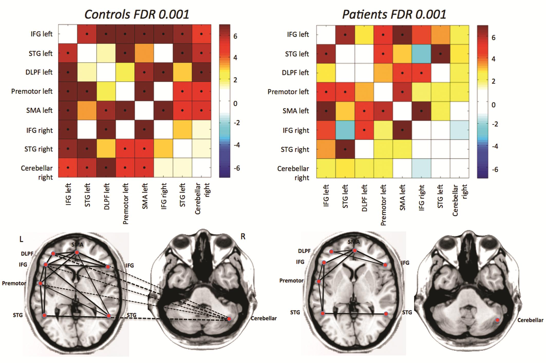
Fig. 1: Differences in functional connectivity from rfMRI between autistic patients and age- and gender matched controls: the major disconnection is between the cerebellum and frontal language areas.

Fig. 1: Differences in functional connectivity from rfMRI between autistic patients and age- and gender matched controls: the major disconnection is between the cerebellum and frontal language areas.

Read more…