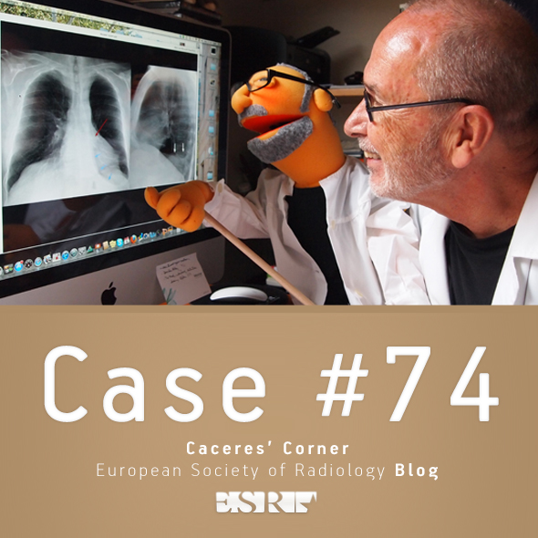
Dear Friends,
Today I’m presenting radiographs of a 54-year-old male with chest pain (below).
Would you call these radiographs normal?
If not, where is the abnormality?
Leave me your answers in the comments section and look out for the answer on Friday.
Read more…

Dear friends,
This week Muppet and I are going on a dangerous trip abroad. Forgive me if I answer your comments a little bit later than usual; the locals may be hostile and refuse to share their WiFi.
Radiographs belong to a 73 y.o. male with fever and symptoms of acute prostatitis.
How do you interpret the chest findings?
Read more…

Dear Friends,
To continue reviewing basic chest patterns, I present the case of a 52-year-old man with a solitary pulmonary nodule.
Diagnosis:
1. Probably malignant
2. Probably benign
3. Indeterminate
4. Need previous films
Check the images below, leave your thoughts and conclusion in the comments section and come back on Friday for the answer.
Read more…

Dear Friends,
The Muppet is very happy with your responses to the previous case. As a prize, he wants to show you the following case that we saw together this July before he went on vacation with Miss Piggy (and left me working). Radiographs belong to a 46-year-old woman, with pain in the chest.
Leave your thoughts and diagnosis in the comments section below, and come back on Friday for the answer.
Diagnosis:
1. Empyema necessitatis
2. Chest wall tumour
3. Metastatic disease
4. None of the above
Read more…

A-236 Role of tomosynthesis in lung imaging
M. Båth | Saturday, March 9, 08:30 – 10:00 / Room G/H
Tomosynthesis is an imaging technique that in recent years has become available for lung imaging. Using low-dose projections of the chest, acquired over a limited angular range, an arbitrary number of section images can be reconstructed, enabling the chest to be visualised in millimetre-thick slices at a very low effective dose. Compared to conventional chest radiography, the disturbance of overlapping anatomy (the main limiting factor for detection of pathology, e.g. pulmonary nodules in chest radiography) is considerably reduced in chest tomosynthesis. Early evaluations have also shown that the detectability of pulmonary nodules is significantly higher in chest tomosynthesis than in conventional radiography. However, compared to computed tomography (CT) the limited angular range used in tomosynthesis results in a reduction in depth resolution, not allowing tomosynthesis to reach the same detection rate as can be obtained with CT. Especially, pathology in the subpleural region may be more difficult to interpret. Nevertheless, most lesions are visible in retrospect on chest tomosynthesis, suggesting that the technique may be suitable for follow-up. This presentation will summarise early evaluations and reported clinical experiences of the technique, as well as describe some of its strengths and limitations.

Dear Friends,
This year I plan to show only chest cases, emphasising the diagnostic approach to basic patterns in the plain film. Hopefully, this monographic approach will help you with the diploma examination.
Cases will be posted every other Monday and answers will be given on Friday.
Radiographs (below) of the first case belong to a 57-year-old man, asymptomatic. Study them carefully, leave your opinions in the comments section, and look out for the answer on Friday.
Diagnosis:
1. Thymoma
2. Teratoma
3. Mediastinal fat
4. Can’t tell
Read more…

Dear friends,
After a two-month vacation, we return with renewed energy. I had a long conversation with Dr. Pepe and we decided to present our cases alternately: I will show my cases one week and Dr. Pepe will present the diploma cases the following week. Cases will be posted on Monday morning and answers will be offered on Friday. This way we will not compete with each other and can remain good friends.
Considering that most of you are still on vacation or with a half-functioning brain, Muppet has selected an easy warm-up case of a 46-year-old woman, asymptomatic.
As usual, leave your thoughts and answers in the comments section below.
Diagnosis:
1. Filariasis
2. Talc aspiration
3. Cysticercosis
4. None of the above
Read more…

Dear Friends,
Presenting the last case before Summer vacation. Muppet needs quality time to replenish his energy (July and August) and will return in September with more interesting (and teaching) cases.
These radiographs belong to a 52-year-old male with a cough. The answer has now been added, but you can still leave your thoughts and diagnosis in the comments section, below, before you check it.
Diagnosis:
1. Mediastinal fat
2. Dilated SVC
3. Lymphoma
4. None of the above
Read more…

Dear Friends,
Showing you the last case before Summer vacation (July and August). Presenting radiographs of a 47-year-old man with moderate dyspnea.
Diagnosis:
1. Collapse of right lung
2. Right pneumonectomy
3. Congenital absence of right lung
4. None of the above
Read more…

Dear Friends,
This week we’re presenting another case provided by my friend Jose Vilar: a 39-year-old woman with cough and moderate dyspnea.
Diagnosis:
1. Mediastinal fat
2. TB
3. LUL collapse
4. None of the above
Read more…


