Dear Friends,
My former disciple Jordi Andreu has contributed this week’s case. Below is a pre-operative PA chest radiograph of a 27-year-old male with a growing perineal mass.
Diagnosis:
1. SVC obstruction
2. Coarctation of aorta
3. Lymphoma
4. None of the above
Read more…

Dear Friends,
Muppet is running out of chest cases and wishes to show a bone case instead. Presenting an AP radiograph of both hands of a 59-year-old male with pain in the joints.
Diagnosis:
1. Rheumatoid arthritis
2. Psoriatic arthritis
3. Hyperparathyroidism
4. None of the above
Read more…

Dear Friends,
This week I’m showing a new ‘Face the Examiner’ case. Presenting the radiographs of a 49-year-old woman with dyspnea.

Dear Friends,
Today we’re presenting a routine chest control of a 53-year-old woman who had a lumpectomy for carcinoma of the breast three years ago. Radiographs were read as normal. Do you agree? Any ideas?
Read more…
Dear Friends,
Today, we’re showing you radiographs of a 29-year-old non-European male with moderate dysphagia. Questions:
1. Where is the lesion?
2. What would be your diagnosis?
Read more…

Dear Friends,
Muppet insists on presenting another case of inspiration/expiration films to test your knowledge. Showing radiographs of a 29-year-old asymptomatic female. What do you think is happening?
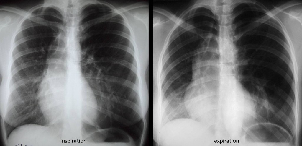
29 y.o. asymptomatic female
Click here for the answer to case #59
Findings: PA radiographs in inspiration/expiration show hyperlucency of the left lung. The heart is displaced towards the right and there is shifting of the mediastinum towards the right on expiration, confirming air trapping of the left lung. In addition, a tubular structure is visible in the left lower lung (arrow).
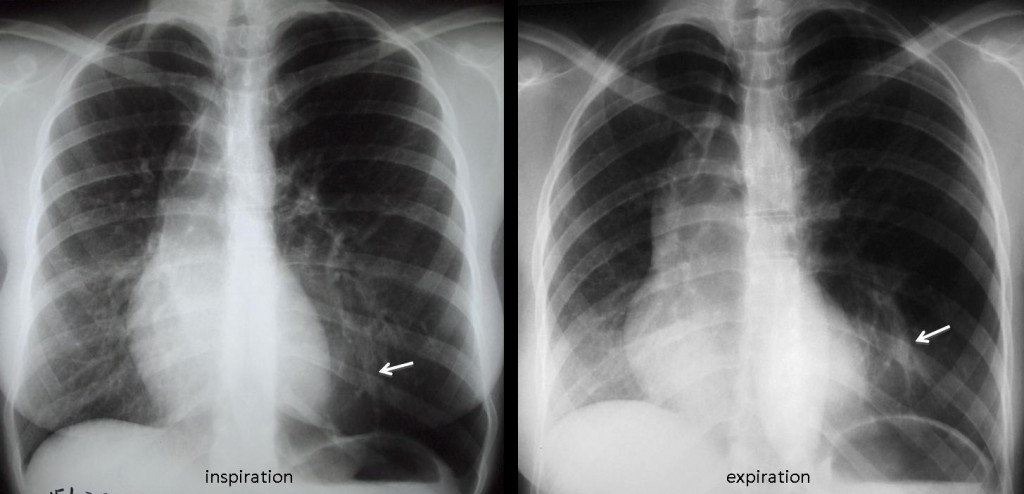
Axial and coronal CT show hyperlucency of the left lower lung, limited by normal upper lung (white arrows). There is a non-enhancing tubular structure in the centre of the left lower lung (red arrows). The displacement of the heart towards the right is due to the expanded left lung plus a marked pectus excavatum.
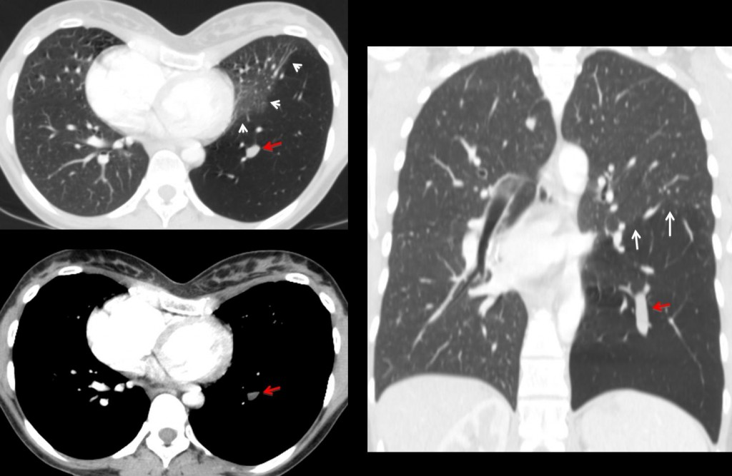
Final diagnosis: Congenital bronchial atresia, left lower lobe.
Teaching point: Congenital lung lesions are not rare in adults. Focal hyperlucent lungs with air trapping and mucous impaction are findings highly suggestive of bronchial atresia.

Dear Friends,
While I was in Vienna, Muppet prepared the following case for you: 21-year-old girl with marked dyspnea, no fever. What do you think it’s happening?
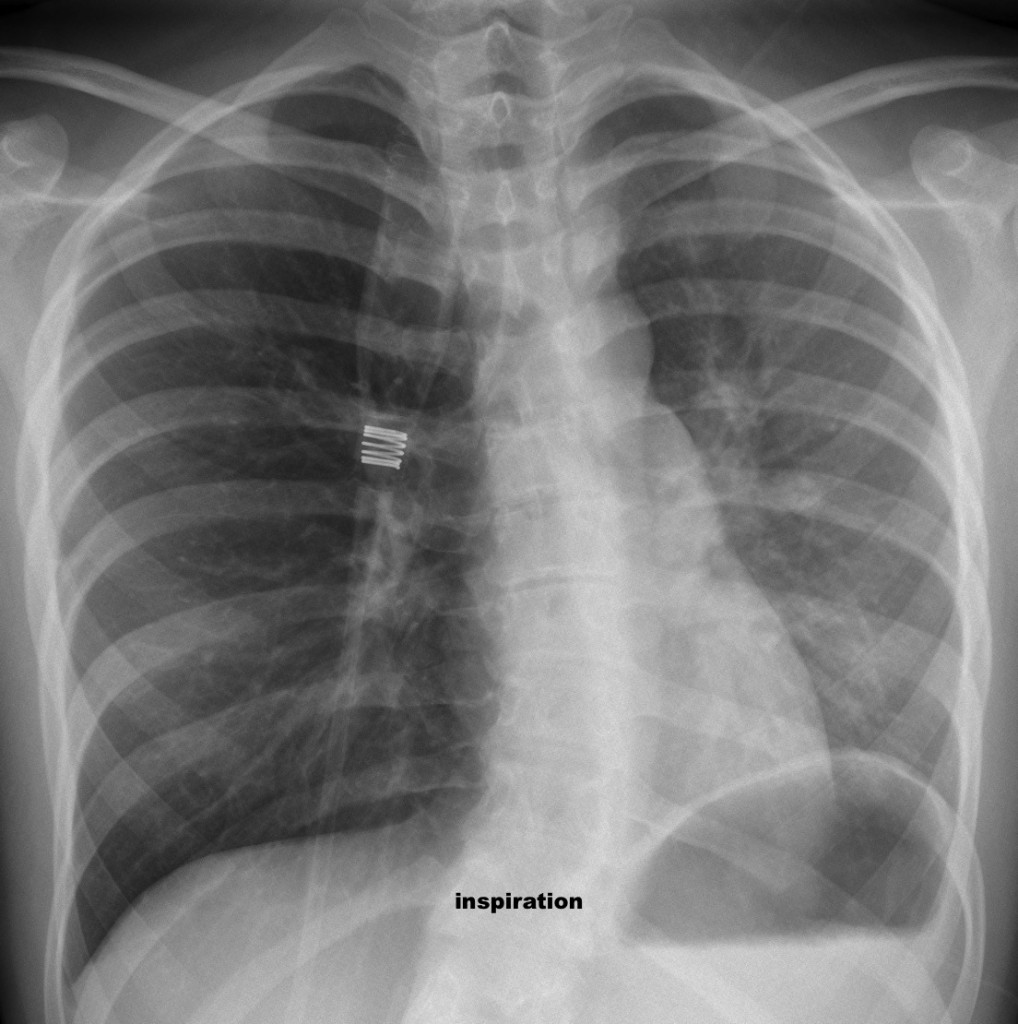
Inspiration – 21-year-old girl with marked dyspnea, no fever.
Read more…
Dear Friends,
Dr. Manel Martínez, a long-time friend of the Muppet, has provided the following case: an asymptomatic 67-year-old man in whom a mass was discovered in the chest radiographs.
Diagnosis:
1. Aortic aneurysm
2. Hydatic cyst of lung
3. Left pulmonary artery aneurysm
4. None of the above
Read more…
Dear friends,
Muppet cannot find difficult cases and, despite himself, is forced to show easy ones. Today we’re showing radiographs of a 48-year-old woman who has had moderate dyspnea for a while.
Diagnosis:
1. Swyer-James syndrome
2. Tumour left main bronchus
3. Old TB left lung
4. None of the above
Read more…

Dear friends,
This week I’m presenting you another new ‘Face the Examiner’ case which simulates a real examination. Showing radiographs of a 35-year-old male with high fever and left pleuritic pain.


