ECR 2019 Cases of the Day Quiz Winners

The Cases of the Day Quiz featured 20 challenging cases covering different sections of radiology.
The cases were shown during ECR 2019 and attendees were encouraged to submit their diagnosis.
From the 374 participants, the following solved most cases correctly:

Elliott Rees; Sawston, Cambs/UK
Filip Vanhoenacker; Antwerp/BE
Ümit Tüzün; Istanbul/TR
Artur Komorowski; Owczary/PL
Manabu Minami; Yokohama/JP
Jan Balak; Prague/CZ
Martina Slamova; Nove Mesto Na Morave/CZ
Diogo Roriz; Coimbra/PT
Veronika Pesti; Budapest/HU
Timothy Sadler; Cambridge/UK
Miguel Correia da Silva; Porto/PT
Vendoti Nitheesha Reddy; Bengaluru/IN

To view the cases of ECR 2019 please click here. 

Congratulations to all winners!

20
Mar 2019
POSTED BY
POSTED IN ,
DISCUSSION 0 Comments

Welcoming all radiographers to ECR 2019

By Jonathan McNulty, EFRS President

Radiographers have long been involved in the European Congress of Radiology (ECR), and the European Federation of Radiographer Societies (EFRS) has had responsibility for developing the radiographers’ sessions since ECR 2012. However, it is only in recent years that the ECR has become the official scientific congress of the EFRS, and the European Society of Radiology (ESR), for medical imaging radiographers.

Over the past eight congresses, the radiographers’ programme has grown considerably, as has the participation of radiographers. A total of 2,177 radiographers and radiography students, from 75 countries, attended ECR 2018 and we look forward to welcoming even more to ECR 2019, which has now become one of the largest international gatherings of radiographers.
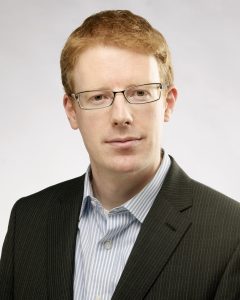
Dr. Jonathan McNulty is Associate Professor and Associate Dean of Graduate Studies at University College Dublin School of Medicine in Dublin, Ireland, and President of the European Federation of Radiographer Societies.

In 2018, a total of 22 refresher courses, professional challenges sessions, special focus sessions, joint sessions, Rising Stars sessions, MyT3 sessions, and scientific sessions made up the radiographers’ programme. For ECR 2019, this will rise to 28 sessions, which will truly offer something for everyone. A special word of thanks must go to Dr. Andrew England from the University of Salford, UK, and a member of the EFRS Educational Wing Management Team, and Dr. Maríanna Garðarsdóttir from Landspitali University Hospital, Iceland, who are the co-chairs of the 2019 radiographers’ scientific subcommittee, and to their team for an excellent educational and scientific programme. Aside from the above sessions, we also look forward to the radiographers’ Voice of EPOS sessions, the involvement of radiographers in a series of sessions at the Cube 2.0 (a special programme dedicated to interventional radiology), and the EFRS Educational Wing annual meeting and our student session.

At ECR 2019, Room C on the 2nd level will become the new venue for most of the sessions in the radiographers’ programme. The Radiographers’ Lounge has also been relocated to Foyer C (outside Room C). In this area, the EFRS and ESR will welcome 20 national radiographers’ societies, along with some educational institutions, who are members of the EFRS Educational Wing, who will all have booths in this area. The radiographers’ Voice of EPOS stage will also be located in the lounge area, as will a number of research studies, requiring your participation, which will take place in the EFRS Radiographers’ Research Hub (Room 2.96). The Radiographers’ Lounge will thus be a great meeting place and, together with the rest of the EFRS Executive Board, I look forward to meeting you in this area and seeing you at the radiographers’ sessions. Read more…

Welcome to ECR 2019!

Welcome to the 25th ECR in Vienna!

Thanks for coming to be a part of ‘the bigger picture’!


By Lorenzo E. Derchi, ESR President

ECR 2019 marks an important anniversary. This will be the 25th ECR congress held in Vienna since 1991: a great achievement! Those who have been coming here since the beginning will find a meeting that has become bigger over the years, now including not only the Austria Center, but also many of the buildings surrounding it.

It is a true ‘congress city’, which has grown to accommodate the increasing numbers of delegates from all over the world. Those who are here for the first time will discover a congress with a wide range of opportunities to learn about the different aspects of our specialty, with sessions suited to all, from beginners to advanced professionals.

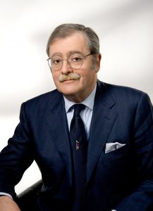
ESR President Prof. Lorenzo E. Derchi is Head of the Department of Radiology at the University of Genoa, Italy.

Over the years, the ECR has provided radiologists with a European platform for the presentation of research and debate and has been the key to the creation of a community of people working together and advancing in science and education together. This has been the result of the efforts of 24 Congress Presidents and Programme Planning Committees who, with the help of the well-structured and dedicated efforts of the ESR Scientific Programme Department, have worked as in a relay team, passing the baton from one to the next and ensuring continuity of aims and good results over the years.

The ECR has always been a creative meeting. It started with a ‘classical’ structure, made up of refresher courses, scientific presentations and posters, and, over the years, has incorporated various new ways for contributors to present scientific work. The first big innovation was in 2003, with the introduction of the electronic posters of EPOS. Then, in 2016, came the new session format ‘the Voice of Epos’ in which the scientific message of selected posters could be explained in person by their authors. Last year, the ‘MyT3’ presentations, the lounge meetings of ‘Coffee & Talk’ and the hands-on approach to interventional radiology of ‘the Cube’ were introduced. All these session formats have proven successful and will be continued, and even expanded, this year. Furthermore, an additional new session type will be launched, not only for ECR delegates, but also open to anyone who is interested in joining in. The ‘Women in Focus’ initiative will explore the challenges encountered by women in our field and in other medical and medical-related professions. It will open a debate over gender parity in our profession and will try to present role models to whom the ever-increasing number of women entering into radiology can relate. Read more…

Russia’s preeminent radiologist continues to reach for the top, after two decades with the ESR

Professor Valentin Sinitsyn is chair of radiology and head of the radiology department at the medical faculty of Moscow Lomonosov State University, Moscow. A pioneer in cardiac imaging in Russia, he has helped advance the field in his country, but believes there is still much to be done to increase MR and CT use. After 20 years of working to advance the ECR and European radiology, he will receive the ESR Gold Medal today.

Sinitsyn has witnessed the introduction of spiral and multidetector CT, high-field MRI and hybrid imaging, and the development of evidence-based radiology and medicine.

Professor Valentin Sinitsyn from Moscow

Professor Valentin Sinitsyn from Moscow, a pioneer in cardiac imaging in Russia, will receive the ESR Gold Medal today.

As he graduated from Sechenov Medical University in Moscow in 1984, cardiac CT and MRI did not yet exist. “With the help of these new modalities, radiology penetrated fields where it had never been used before, and the best example is cardiac imaging,” he said.

Sinitsyn started implementing cardiac imaging back in the 1980s and has very fond memories from that time. Although these were also harsh days for the field, he said.

“Most cardiologists and radiologists believed that cardiac MRI and CT were toys that were good for nothing in real clinical practice. I still remember my first case of apical hypertrophic cardiomyopathy diagnosed with cardiac MRI in 1987, when nobody could make a correct diagnosis,” he said.

Today he admits to being a little disappointed with the slow penetration of cardiac MRI and CT in Russian clinical practice. The benefits of modern cardiac imaging are well known, but the percentage of cardiac CT and MR examinations in cardiac imaging is just around 1%. “Low reimbursement for such examinations and insufficient knowledge and motivation from both radiologists and cardiologists are mostly to blame for this situation,” he said.

Read more…

Win FREE registration for ECR 2019 via Twitter!

10,000 twitter followers?! This is a reason to celebrate!

We have THREE FREE places at ECR 2019 to give away to our fantastic followers.

To be entered into our draw, all you need to do is:

1) Follow the European Society of Radiology’s Twitter account, @myESR (existing followers also qualify)

2) Either like, comment, or re-tweet one of our tweets between now and June 10 (deadline: 23:00 CEST).

All personal* Twitter accounts meeting those two simple criteria will be entered into our random draw for free ECR 2019 registration.

Thanks for following us and good luck!

*Accounts representing companies, societies, institutions or other organisations will not be eligible. Anyone found to use multiple accounts to enter will also be ineligible.
Full terms and conditions of this competition can be found here.

ECR 2018 Cases of the Day Winners

The winners of the ECR 2018 Cases of the Day Quiz are as follows:

Mehmet Serindere; Hatay/TR
Sungeun Park; Seoul/KR
Seungchul Han; Seoul/KR
Sewoo Kim; Seoul/KR
S. A. Sohaib; Sutton/UK
Rafal Darecki; Koscierzyna/PL

To view the cases please click here.

Congratulations to all winners!

26
Mar 2018
POSTED BY
POSTED IN ,
DISCUSSION 0 Comments

The best submissions for the “Normal variant or disease” interlude at ECR 2018

Dear Friends,

Over the last couple of years, one of the last sessions at the ECR has always covered 20 interesting cases from various subspecialties, which the audience is asked to solve in an interactive way to broaden and update their knowledge.

In between, the very best submissions from the global radiological community have been presented in an interlude lecture. The best submission has always been awarded with a prize and a certificate.

Due to time limits, not all submitted cases can actually be shown onsite, but the session’s rising popularity has resulted in increasing numbers of submissions of excellent quality and didactic value. This is why we would like to give our submitters the opportunity to reach a broader audience by posting the best cases here on the ESR Blog.

This year’s topic was Normal variant or disease to avoid misdiagnosis, overdiagnosis, unnecessary and costly work-up, as well as patient’s concerns.

When we think back of our time in medical school and remember our studies of embryology, we have probably all thought at some stage, how astonishing it is that a viable fetus develops and a healthy newborn is delivered. Embryological development is so complex that an awful lot can go wrong at any stage. Fortunately, real malformations are rare. In our professional life as radiologists, though, we are not infrequently dealing with normal variants, when development is arrested at a certain stage or turns the wrong way round. Even first year residents are familiar with right sided aortic arch, azygos lobe and accessory ossification centres. Over time, one encounters a whole array of normal variants and learns to interprete them as such. Of course, one can only identify, what one has learnt to see and there is a large spectrum of slight developmental abnormalities, which should not be misdiagnosed as abnormality in need of further imaging, invasive diagnostic procedures or treatment. On the other hand, some developmental disorders have clinical significance and you could be the first to advise the patient.

Below, we are pleased to present the best submissions for the “Normal variant or disease” interlude at ECR 2018:

Incoming ESR President Lorenzo Derchi looks forward to celebrating an ECR milestone in 2019


By Julia Patuzzi

As has been our tradition for a few years now on the final day of our congress, we already look ahead to next year’s ECR. We therefore spoke with Prof. Lorenzo E. Derchi from Genoa, Italy, who, as incoming ESR President, is in charge of ECR 2019. He told us about his plans and ideas for the next European Congress of Radiology.

ECR Today: Professor Derchi, next year’s European Congress of Radiology will be the 25th to be held in Vienna. Are there any specific celebrations planned that you can already share with us?

Prof. Lorenzo E. Derchi

As incoming ESR president, Prof. Lorenzo E. Derchi, Head of the Department of Radiology at the University of Genoa, will preside over ECR 2019.

Lorenzo E. Derchi: We are still thinking about them. An anniversary like this is something more than an opportunity for a big celebration. It is the right moment to re-think our history and what we have reached over the years. To always have the congress in the same city since 1991, at first every other year and then, after 1999, annually, has been a radical change. The new formula proved successful and ECR has become the European meeting to come to in order to learn the latest clinical and technical advances in our discipline as well as to present the results of the previous year’s research.

In my experience, however, it has become much more. From 1991 up until today all congress chairpersons have been part of the same team in a relay race, each handing off the baton to the next one. This continuity can be felt in the special atmosphere at each meeting, and has allowed the congress to become the annual appointment to meet old friends and make new ones; to discuss any radiological topic and then to start working together on it; and finally a place to meet periodically to share problems, advancements and results of long-term projects.

ECRT: Were you here for the first ECR in Vienna in 1991? If not, when did you attend your first ECR? What do you recall from that visit?

Read more…

Trojanowska to deliver honorary lecture on neglected form of cancer

Polish radiologist Dr. Agnieszka Trojanowska will shed light on human papilloma virus (HPV)-induced squamous cell cancer of the head and neck, a common yet long neglected type of cancer. In an interview with ECR Today, she also explained the origins of her passion for head and neck imaging, and how radiologists will increasingly benefit from computer science.

Dr. Agnieszka Trojanowska, assistant professor in the department of radiology and nuclear medicine at Lublin University Hospital, is an internationally recognised specialist in head and neck imaging. Her special interest for oncology and her natural curiosity led her to agree to deliver the Josef Lissner Honorary Lecture on HPV-induced squamous cell cancer of the head and neck (HNSCC) at ECR 2018.

Agnieszka Trojanowska from Lublin, Poland, will speak on HPV-induced squamous cell cancer of the head and neck in her Honorary Lecture today.

“HNSCC behaves differently than more well-known forms of SCC, and it is becoming more and more prevalent. It affects young, well-educated people with high socioeconomic status, and is a quickly rising sexually transmitted entity with peculiar clinical and molecular characteristics. In particular the rise of HPV-induced cancer has been observed in the United States since the beginning of 21st century,” she said.

It was important for her to talk about this long overlooked topic, for which much remains to be done. “It has been under-estimated for many years in many countries. Nowadays, we can speak about an HPV epidemic, leading to a significant rise of oropharyngeal cancer incidences worldwide. These cancers are quite unique and, in my opinion, knowledge of their physiology and treatment options is essential,” she said.

New research shows that, compared with environmental-related head and neck squamous cell carcinomas, patients with HPV-related malignancies display a better response to treatment and a lower risk of death and progression, Dr. Trojanowska pointed out. It is also noteworthy that, according to the 2017 TNM classification, HPV+ cancers are evaluated with a separate grading scale, she added.

Read more…

Riklund to unveil the next big thing in hybrid imaging

The Swedish radiologist Prof. Katrine Riklund, the current Chair of the ESR Board of Directors, has dedicated her career to the development of hybrid imaging. During todays’ Marie Curie Honorary Lecture (Room A, 1:00 p.m.) she will look back at the achievements made in this emerging field and look forward to future advances.

Riklund, who is a professor, consultant in diagnostic radiology and pro-vice-chancellor of Umeå University, is one of Sweden’s leading radiologists. She is also one of the first researchers to have recognised the potential of combining PET with CT and MRI, and has worked to advance the field ever since its emergence at the beginning of the 21st century.

“The combination of structural and functional/molecular imaging is fascinating. The entire field is new and extremely interesting, and it’s the closest to my heart when it comes to imaging,” she said.

Prof. Katrine Riklund from Umeå, Sweden, will speak about the growing significance of hybrid imaging in today’s honorary lecture.

During her lecture, Prof. Riklund will share examples of what hybrid imaging can do and what is going to come next. A major trend will be making use of the entire hybrid imaging examination for diagnostic protocols, also for the CT part, she believes. “This changes workflow and gives us more information. PET and CT or MR are not competing techniques, they are complementary,” she said.

Tracer development is key for PET but digital detectors will also represent a major step forward. Currently, the need for a cyclotron for production of radionuclides hinders substantial distribution of scanners outside large centres. “To make hybrid imaging really take off, we need other forms of tracer production. I would like to see the tracer production work like a coffee machine – with buttons to select tracer and radionuclide,” she said.

In her day-to-day work, Prof. Riklund is involved in various research projects, such as COBRA, a prospective multimodal imaging study of dopamine, brain structure and function, and cognition; (PEARL-PD), 18F-FE-PE2I PET/CT, a study of dopamine transporters in early Parkinson’s disease, RECTOPET (REctal Cancer Trial On PET-MR/CT); and Prostate Cancer – PSMA and Acetate in PET/CT and PET/MR. These projects reflect her three major interests in the field; movement disorders and cognition in central nervous system, and prostate and colorectal cancer.

Read more…