Interview: Joanna Fairhurst, consultant paediatric radiologist from Southampton, UK

IDOR_2015_Facebook_851x315px_72dpi_RGB

An interview with Joanna Fairhurst, consultant paediatric radiologist at the children’s radiology department of the University Hospitals of Southampton.

European Society of Radiology: What is paediatric imaging? What age are the patients, and how is it different from regular imaging?
Joanna Fairhurst: Paediatric imaging covers all imaging modalities – plain films, ultrasound, fluoroscopy, computed tomography (CT), nuclear medicine, magnetic resonance (MRI) – undertaken in children ranging from new-born infants to those who are sixteen, or in some centres eighteen, years old. Imaging patients in this age range poses some very specific challenges. First, coming to the hospital can be a very frightening experience for young children, and we need to adapt our techniques to help children feel as secure and comfortable as possible, and we often employ distraction and play therapy to reduce their anxiety and help them cooperate with their examinations. We also try to create a child-friendly environment, by decorating our department and providing toys, but the best way to make our young patients feel at ease is to have radiographers and radiologists who are experienced in, and enjoy working with children.

Joanna Fairhurst is a consultant paediatric radiologist at the children’s radiology department of the University Hospitals of Southampton

Joanna Fairhurst is a consultant paediatric radiologist at the children’s radiology department of the University Hospitals of Southampton

The next main difference between paediatric and adult radiology is that we have to be familiar with the imaging appearances of the developing patient – from pre-term infant to adolescent – including many normal developmental variants. We also have to deal with many diseases and pathologies that are specific to children. Unlike other subspecialties within imaging, although some do specialise, most paediatric radiologists are involved with all modalities and all body systems.
Finally, when we image children, we not only have to communicate with young people: very often we also have to interact with their parents and carers, so we must learn to respond to their concerns and needs as well.

ESR: Since when has paediatric imaging been a specialty in its own right?
JF: It could be argued that paediatric radiology is the oldest imaging specialty, dating back to the use of x-rays and the interpretation of the images produced in children’s hospitals in the early 1900s. Many people, however, consider Dr. John Caffey (1985–1978) as the ‘founder’ of paediatric radiology after he published his book Pediatric X-Ray Diagnosis in 1945. Paediatric imaging gained broader recognition in the United States with the founding of the Society for Pediatric Radiology in 1958, and came of age in Europe with the creation of the European Society of Paediatric Radiology in 1963.
Read more…

Reducing kids’ MRI anxiety, brick by brick

guest post by Benjamin Taragin

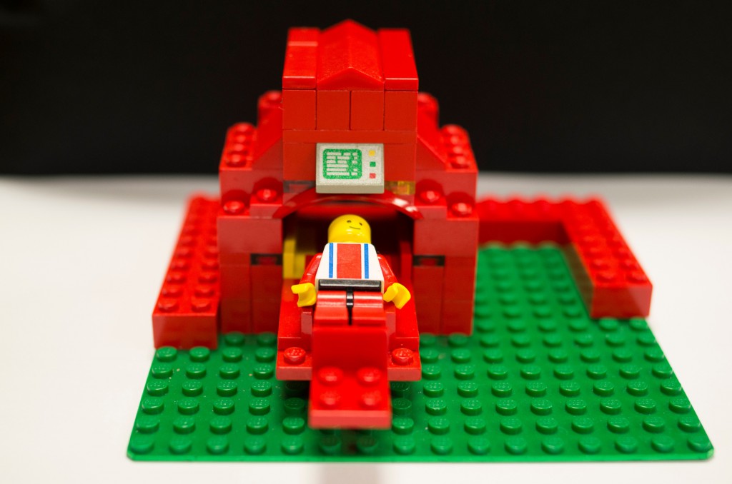
Lego Blog Image 1

I have always been fascinated by LEGO®. There is something soothing about raking the pile with your hands, trying to find the perfect piece to complete your next build. It was with this mindset that I was helping my son, Yoni, build one of his projects (clearly there is no pleasure in this for me ;)). As we were working with the LEGO pieces, I came across a curved semicircular piece and realised that it reminded me of the bore opening of an MRI. With that thought in my head, my son and I began to build our initial LEGO MRI model. After building it, I realised that this might actually be useful for our child life division at the Children’s Hospital at Montefiore, spearheaded by Susan Frank and Meghan Kelly, to use when prepping patients for MRI. While many simulators exist on the market, some large and some small, none are built with the basic blocks of childhood. Additionally, its small size and portability allows it to be carried around the hospital in a regular work bag.

Read more…

Caceres’ Corner Case 123 (Update: Solution)

ESR_2015_Blog-CaceresCorner-123

Dear Friends,

To mark the International Day of Radiology, which this year is focusing on paediatric imaging, we are presenting chest radiographs of a 5-year-old boy with an abdominal mass and mild fever. A Wilm’s tumour was suspected. Check the images below, leave your thoughts in the comments section and come back for the answer on Friday.

Diagnosis.

1. Pleural tumour in fissure
2. Pneumonia
3. Metastases
4. None of the above

Read more…

American and European emergency radiologists compare experiences

ECR2014_ECRToday_Blog_Final

Watch this session on ECR Live: Friday, March 7, room F1
Four separate sessions, starting 08:30, 10:30, 14:00, and 16:00
Tweet #ECR2014F1 #MC422 #MC522 #MC622 #MC722

For the fourth year in a row, the European Society of Radiology (ESR) and the Radiological Society of North America (RSNA) will hold a joint course on a hot topic in radiology. After oncologic imaging for the past three years, emergency radiology will be under the scope of North American and European radiologists, who will share and compare their experiences in the dedicated Mini Course today at the ECR*.

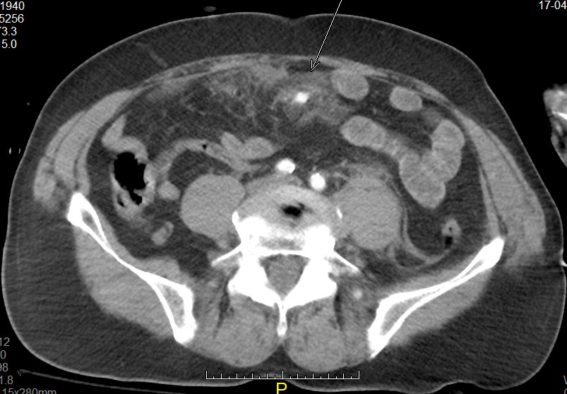
MDCT is the best modality for identifying lesions as subtle as active mesentric bleeding and infiltration (arrow) caused by blunt abdominal injury (lap-belt compression). (Provided by András Palkó)

MDCT is the best modality for identifying lesions as subtle as active mesentric bleeding and infiltration
(arrow) caused by blunt abdominal injury (lap-belt compression). (Provided by András Palkó)

Medical emergencies and trauma are responsible for high levels of morbidity and mortality in all age groups worldwide, and trauma is the leading cause of death in people younger than 45. Consequently, the use of imaging in emergency departments has been increasing exponentially since the 1990s. The demand for CT examinations from emergency departments is, for instance, growing annually by 5% to 15%, depending on the institution.

Even with all the advanced tools imaging has to offer, trauma remains a challenge for most emergency services, according to Ulrich Linsenmaier, associate professor of radiology and head of the department of diagnostic and interventional radiology at KMPP Clinics in Munich, Germany. “Even for advanced level 3 medical centres running their own emergency radiology unit, it is a challenge to integrate advanced radiology services into an interdisciplinary team treating patients with acute traumatic and non-traumatic emergencies,” he said.

Read more…

07
Mar 2014
POSTED BY
POSTED IN ,
DISCUSSION 2 Comments

ECR 2013 Rec: Imaging features of acinar cell cystadenoma: can we differentiate them from branch duct IPMNs? #B0576 #SS901

ECR2013Rec_Blog_Cover_SS901_B0576

B-0576 Imaging features of acinar cell cystadenoma: can we differentiate them from branch duct IPMNs?

C. Delavaud, G. D’Assignies, J. Cros, P. Ruszniewski, P. Hammel, A. Couvelard, V. Vilgrain, M.-P. Vullierme

Purpose: Acinar cystic cystadenoma (ACC) of the pancreas is a rare benign entity first described in 2002, defined by histological criteria. Radiographic appearance had almost not been described so far. Most of the patients underwent surgical resection under the preoperative diagnosis of intraductal papillary mucinous neoplasms. The aims of this study are to define imaging diagnostic criteria of ACC based on radiopathological confrontation and to compare clinical, biological and imaging data between patients with ACC and with branch ducts IPMN.
Methods and Materials: All patients with ACC who underwent pancreatic surgery for suspicion of IPMN and the 20 last patients with histologically proven branch ducts IPMN were retrospectively included. Clinical and biological information were collected from the medical reports. Radiological and histological documents were reviewed in order to define imaging diagnostic criteria of ACC. Data were compared using the Chi-square test or Fisher’s exact test.
Results: ACC was symptomatic in all but one patient. There were no statistical difference between ACC and IPMN group with regard to clinical and biological data. Combination of four radiological criteria allowed differentiating ACC from IPMN: cyst calcification, presence of more than 5 cysts, clustered peripheral small cyst, and absence of communication with main pancreatic duct. Sensibility and specificity were, respectively, 75 and 100 % with combination of at least three of these criteria.
Conclusion: ACC is a rare benign pancreatic tumour with specific imaging features despite some similarities with IPMN. Recognition of this entity may help us to propose the diagnosis and prevent extensive surgery.

ECR 2013 Focus: Justifying CT in Paediatric Radiology

ECR2013_Focus_PaediatricRad

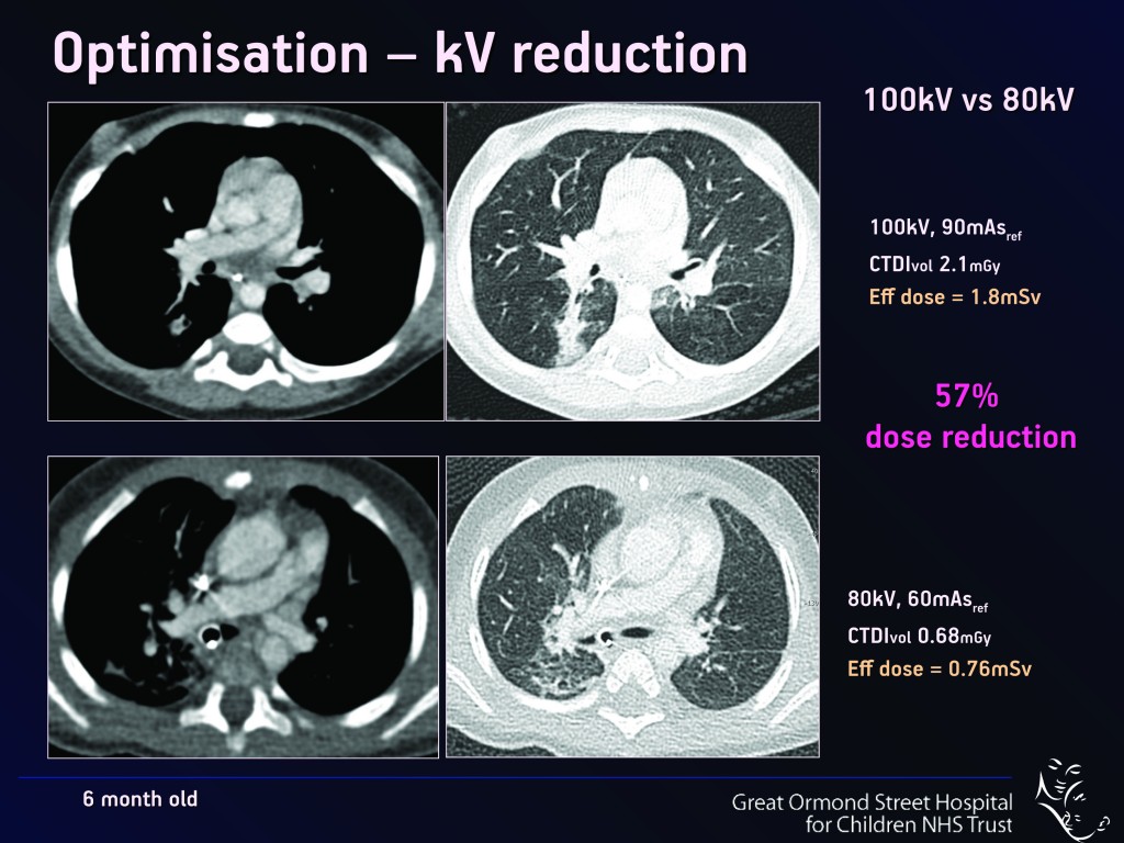
The optimisation and justification of procedures is vital when using CT as an imaging modality; particularly on children, who are more sensitive to ionising radiation than adults. Therefore, it is crucial that all those who use CT understand the physics behind the equipment and ultimately use this understanding to minimise the potential risks while maximising the potential benefits to each individual patient. Patients should also be informed of the risks and benefits of undergoing a CT scan. World-renowned experts will explain these issues in detail during a Special Focus Session at ECR 2013.

“Not all radiologists and technicians are aware of the latest dose reduction strategies. Some are not necessarily so well-informed and perhaps do not realise how important this is. We believe that it is a question of trying to get everybody to a certain level of knowledge and expertise,” said Dr. Catherine Owens, paediatric radiologist and CT unit lead at Great Ormond Street Children’s Hospital in London, U.K.

Paediatric MDCT  Chest Imaging Techniques

Fig. 1

Read more…