
Dear Friends,
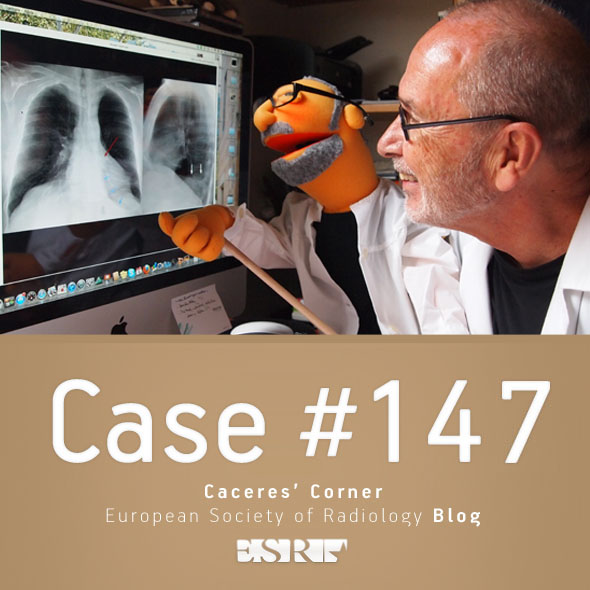
Today we are moving to a new chapter in the Painless Approach to Interpretation, addressing what to do when the chest radiograph does not show an obvious abnormality.
For this purpose I am presenting the PA and lateral radiographs of a 57-year-old man with a chronic cough. What do you see?
Examine the image below, leave your thoughts in the comments section and come back on Friday for the answer.
Read more…
Dear Friends,
Today we are showing a case in recognition of the International Day of Radiology, which takes place tomorrow.
Below are images of a 35-year-old woman with chest pain and progressive dyspnoea for the last three weeks. Leave your thoughts in the comments section and come back on Friday for the answer.
Most likely diagnosis:
1. Lymphoma
2. Pleural metastases
3. Mesothelioma
4. Any of the above
Read more…

Dear Friends,
Now that we’ve looked at the three key questions to ask when facing a chest radiograph (chapters 1, 2 and 3), we move on to the interpretation of pulmonary lesions.
Today I am showing chest radiographs of a 31-year-old woman with marked dyspnoea for the last three days.
What would you call the predominant pattern?
1. Reticulonodular
2. Septal
3. Air-space disease
4. None of the above
Check the images below, leave your thoughts in the comments and come back on Friday for the answer.
Read more…
Dear Friends,
Today we’re showing chest radiographs of a 57-year-old man with chest pain and dyspnoea.
Check the images below, leave your thoughts in the comments, and come back on Friday for the answer.
Diagnosis:
1. Carcinoma of the lung
2. Thymoma
3. Lymphoma
4. None of the above
Read more…

Dear Friends,
Today we are presenting a recent case. The PA radiograph was made for routine check up of a 49-year-old male.
Check the image below, leave your thoughts in the comments and come back on Friday for the answer.
Diagnosis:
1. McLeod syndrome
2. Changes post-TB
3. Congenital lung hypoplasia
4. None of the above
Read more…

Dear Friends,
I hope you remember the three questions to ask when facing a chest radiograph:
a) Is there any visible abnormality? (Chapter 1)
b) Is it intra or extrapulmonary? (Chapter 2)
c) What does it look like?
To discuss the third question, I’m showing chest radiographs of a 36-year-old woman with chest pain.
What does the lesion look like?
1. Pericardial fat pad
2. Thymic tumour
3. Pericardial cyst
4. Any of the above
Check the two images below, leave your thoughts in the comments section and come back on Friday for the solution.
Read more…

Dear Friends,
Today we are showing another pre-op case, including a PA chest radiograph of a 62-year-old man with lumbar hernia. Check the image below, give us your thoughts in the comments, and come back on Friday for the answer.
Diagnosis:
1. TB
2. Carcinoma
3. Pulmonary hypertension
4. None of the above
Read more…

Dear Friends,
As you may remember from Diploma Case 94, when I’m facing a chest radiograph I start by asking three questions:
a) Is there any visible abnormality?
b) It is intra- or extrapulmonary?
c) What does it look like?
Today we will discuss the second question, showing chest radiographs in two different patients. Is the abnormality intra- or extrapulmonary? Check the images below, leave your thoughts in the comments and come back for the answer and discussion on Friday.
Read more…

Dear Friends,
Today we are presenting pre-op radiographs for inguinal hernia in a 37-year-old-man. Check the images below, leave your thoughts in the comments section, and come back on Friday for the answer.
Diagnosis:
1. Pulmonary arterial hypertension
2. Thymoma
3. Pericardial cyst
4. None of the above
Read more…

Dear Friends,
This year I intend to discuss the basic principles of interpreting chest radiographs, under the heading, “The Beauty of Basic Knowledge”.
I plan to structure the discussion into three main parts, which will take us through the whole academic year:
Part 1. A painless approach to interpretation
Part 2. To err is human: how to avoid slipping up
Part 3. The wisdom of Dr. Pepe
This week’s case is the first chapter of my ‘painless approach to interpretation’. Interpreting chest radiographs is not difficult if we follow Confucius’ saying: “ A journey of a thousand miles begins with a single step”. As a clinical radiologist, my first step is to ask myself three questions:
a) Is there any visible abnormality?
b) It is intra or extrapulmonary?
c) What does it look like?
Today I’ll discuss the first question. Below you can see the chest radiographs of three different patients. Do you see any visible abnormality in any of them? Let me know in the comments section and come back on Friday for my answer.
Read more…


