A-339 Chairman’s introduction
M. Krokidis, A.A. Hatzidakis | Saturday, March 9, 16:00 – 17:30 / Room N/O
There are some basic IR techniques everybody needs to know before starting to treat biliary diseases. Τhese are Percutaneous Transhepatic Cholangiography (PTC) and Percutaneous Transhepatic Biliary Drainage (PTBD). These basic procedures are essential first for opacification and then for getting access into the biliary tree. After accessing the biliary system, decisions must be made about the need for further interventional procedures. Presence of benign or malignant strictures, stones or other kind of disease are leading us to the necessary actions, such as dilatation, stenting or lithotripsy. Dilatation of stenosed parts of the biliary system is performed after negotiation of the stricture or occlusion with help of high-pressure angioplasty balloons of variant sizes. Balloons of 8-14 mm width can be used alone or two parallel to each other. Stenting is rarely needed in benign biliary disease and usually in cases where multiple dilatations are not responding. Percutaneous lithotripsy with or without cholangioscopic assistance is a wide-used technique for clearance of biliary stones or fragments. Extraction balloons or baskets and special lithotripsy devices are commonly used for impacted or large calculi.
A-511 fMRI in epilepsy
N. Bargalló | Sunday, March 10, 16:00 – 17:30 / Room G/H
Functional MRI (fMRI) is a non-invasive tool that is capable to detect the subtle homodynamic changes produced in regional brain activation. The main fMRI clinical application until now is localization and evaluation of brain eloquent areas in surgical planning of brain pathology. fMRI application in epilepsy patients are language lateralization, memory function assessment and localization of ictal and interictal BOLD changes. There are several factors that can influence the results of a fMRI experiment such as the scan noise for the rest condition, the simplicity of the task performance, the monitoring of the experiment during the exam, how to achieve a real baseline condition and the most important, to use the most specific paradigm that would activate the selected brain areas. In practical approach, one must be aware that sometime fMRI studies are applied in paediatric or impaired cognitive epilepsy population when deciding the language or memory paradigm, and is recommended to use multiple and feasibility tasks to assure the results. fMRI for language lateralization is currently used in the clinical practice and provides comparable results to the intracarotid amobarbital test (IAT). In fMRI studies for memory function assessment, results show changes in epileptic patients, but further studies are required to validate this technique to an individual level. A new application is ictal or interictal fMRI with EEG recorded that provide more detailed information about simultaneously electrographic and homodynamic changes in the seizure process, with encouraging results for epileptogenic area localization and propagation patterns.
A-245 Pre-therapeutic radiological evaluation
J. Raupach, O. Renc, P. Hoffmann, J. Zizka; | Saturday, March 9, 08:30 – 10:00 / Room N/O
Endovascular abdominal aortic aneurysm repair (EVAR) was introduced over 20 years ago to primarily treat old and sick patients. Due to technical improvements and satisfactory clinical results of this technology, the number of patients treated with stent-grafts is steadily increasing. There is also tendency to use this therapy for ruptured abdominal aortic aneurysms. Pre-operative assessment of aortic morphology regarding suitability for stent-graft implantation is, therefore, an important challenge for every radiologist now. Main limitation of EVAR is unfavourable anatomy of landing zones and access vessels. Gold standard for EVAR planning is contrast-enhanced CTA. Alternative modality for patients with contraindications for CT, such as renal impairment, is unenhanced MR with steady-state free precession sequence. A number of 2D or 3D reconstructions are generated to provide information about the aneurysm morphology. Dedicated vessel analysis and planning software can be applied. Usually, axial images and thin MPR reconstructions are sufficient in emergent cases. Proper stent selection is a domain of operator and is still matter of his/her experience. The planning procedure can be subdivided into 4 different sections: infrarenal neck, aneurysmal sac, aortic bifurcation and access vessels. There are several critical and rules which must be obeyed during the evaluation process and general radiologists should be aware of. The presentation will review main inclusion and exclusion criteria for EVAR.
A-028 Ultrasound elastography
A. Athanasiou | Thursday, March 7, 16:00 – 17:30 / Room F2
Breast ultrasound elastography provides information about tissue elasticity Young modulus E = s / e, where s is the compression (stress) and e is the deformation (strain) of the tissue. It is a complementary tool to breast ultrasonography, easily performed in clinical practice. Two elasticity modes are currently available: strain imaging, where manual compression is applied to the ultrasound probe and tissue displacement is registered; tissue deformation is then calculated by means of dedicated software providing real-time elasticity images (color- or grey-coded) superimposed on B-mode imaging. This is a qualitative or semi-quantitative mode. Shear wave imaging, where US probe is used to induce mechanical vibrations using acoustic radiation force generating local tissue displacement. This mode provides quantitative information about either tissue displacement velocity or tissue stiffness itself in kPa. Functional information provided by elasticity imaging can be particularly useful for BIRADS 3 or 4a lesions. Various studies indicate that elasticity combined to B-mode imaging can improve breast ultrasound specificity up to 75-88%. False negative findings may be encountered in case of “soft” lesions (mucinous, medullary or cystic carcinomas) or inflammatory cancers. Differentiation between echogenic cysts and homogeneous solid lesions (such as fibroadenomas) can be improved as cystic features are usually specific in elasticity imaging. Iso-echoic lesions such as infiltrating lobular carcinomas may be better delimitated. Lymph-node characterization and microcalcification assessment can be improved, although few data are available and need further validation. 3D elastography is actually in progress and would be useful in monitoring response to neoadjuvant treatment.
Watch this session on ECR Live: Sunday, March 10, 08:30–10:00, Room E1
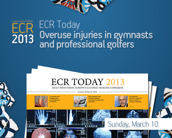
Overuse injuries due to excessive exercise are normally seen in professional athletes, but they are also becoming more frequent in amateur athletes. The Refresher Course on overuse injuries in sports will present three examples of how these injuries, caused by different sports, can be diagnosed and treated.
Gymnastic exercises, for example, are very demanding, not only on the axial but also the peripheral skeleton, and they involve strong forces due to hyperextensive and hyperflexion exercises. A certain degree of hypermobility and increased flexibility is necessary to perform some gymnastic exercises, and so training is required to improve this flexibility. Unnatural movements are sometimes necessary in order to increase this hypermobility and flexibility.
Read more…
Watch this session on ECR Live: Saturday, March 9, 16:00–17:30, Room G/H
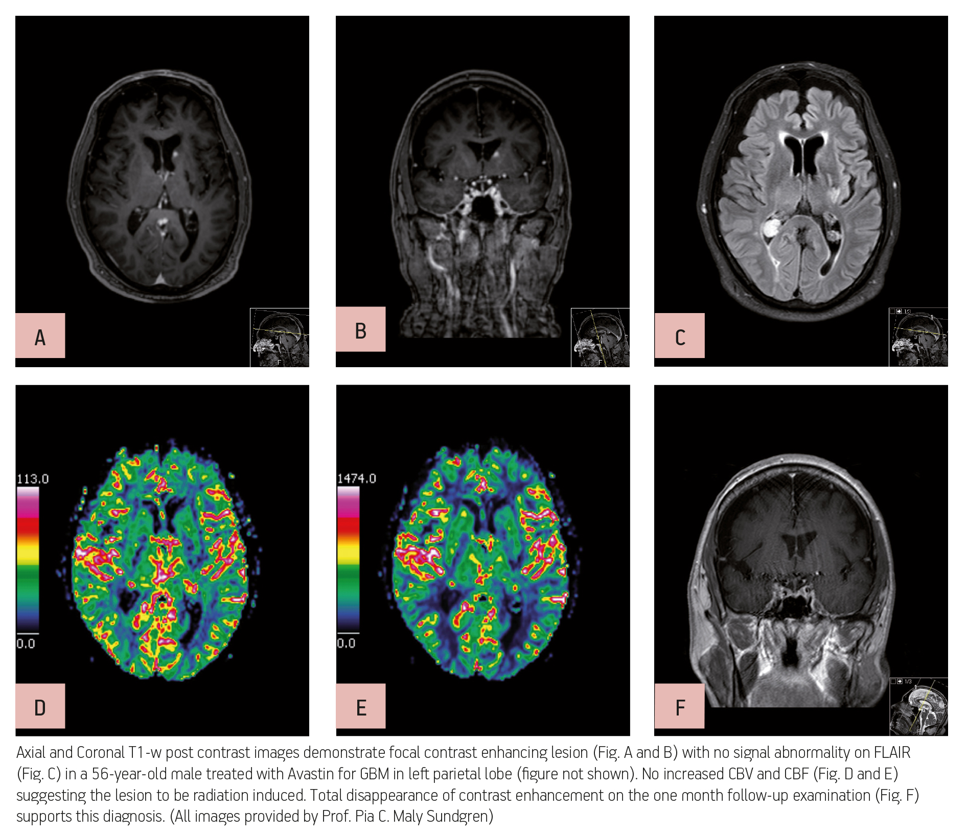
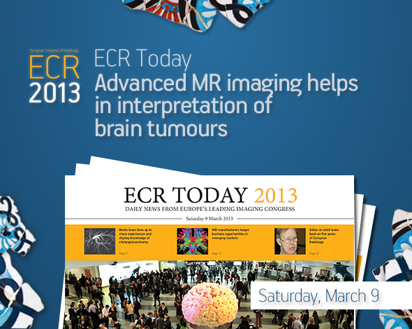
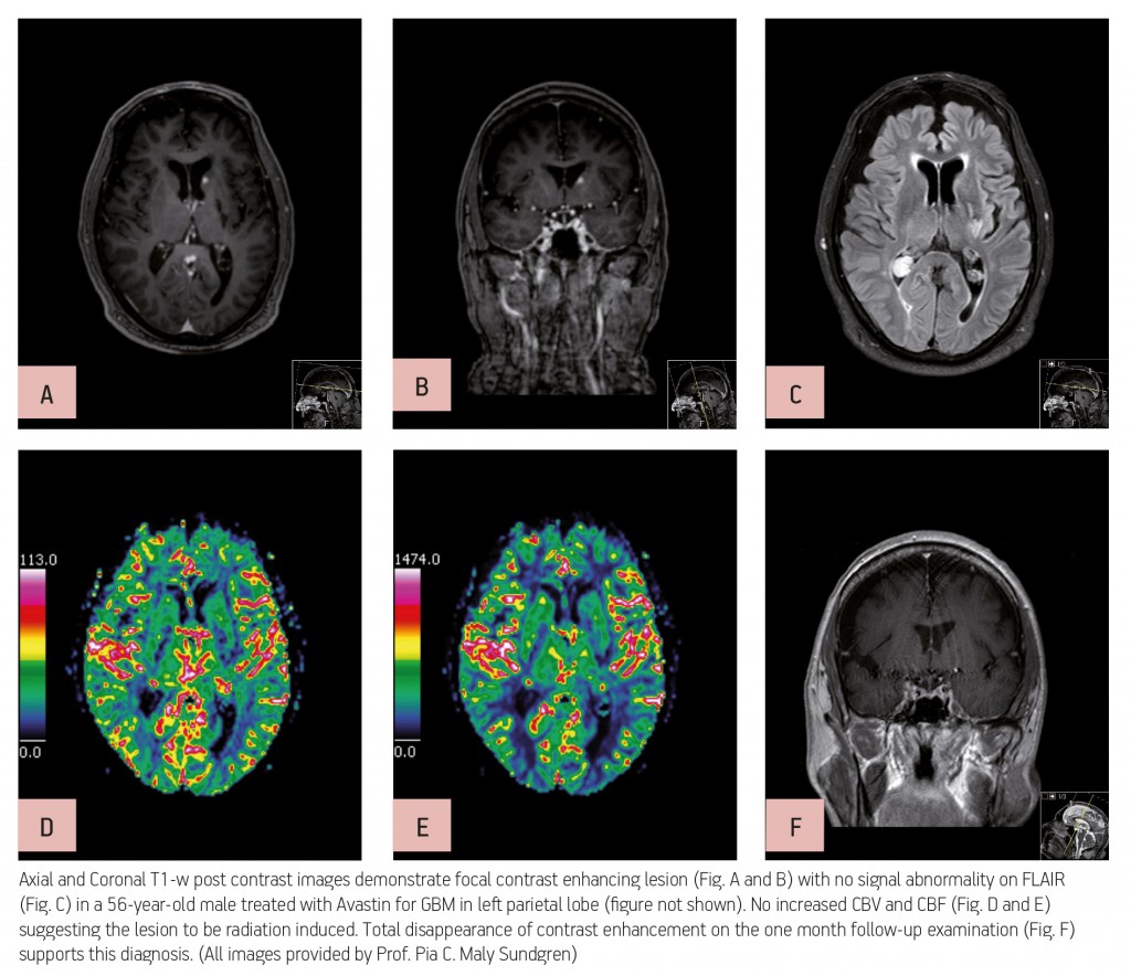
Advanced MR imaging techniques such as perfusion and functional imaging have been a great help in improving the diagnosis and staging of brain tumours. Unlike conventional MR techniques, advanced MR techniques can be used to obtain information not only on the morphological, but also on the functional characteristics of tumours.
One of the most common types of brain tumour is glioblastoma, which is highly malignant and has a high cell reproduction rate due to the fact that it is nourished by a large network of blood vessels. According to the American Brain Tumour Association there are two types of glioblastoma: primary glioblastomas, which tend to form and make their presence known quickly by growing aggressively, and secondary glioblastomas, which are also aggressive but show slower growth and only represent 10% of all diagnoses.
Read more…
Watch this session on ECR Live: Thursday, March 7, 16:00–17:30, Room D2
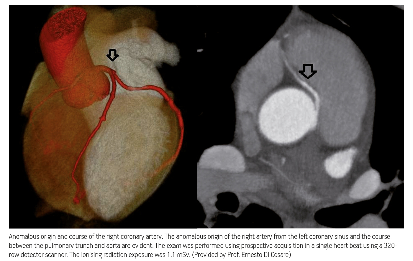
Over the past decade, technological improvements have led to the widespread use of imaging modalities in the prediction, diagnosis and follow-up of coronary disease. Radiologists now have the ability to obtain information on the structure of cardiac muscle with MRI and evaluate cardiac arteries with CT, while hybrid imaging will soon allow them to do both. Cardiac CT will also provide more functional information in the future, and its use will continue to grow. Experts will present the newest and upcoming possibilities of cardiac imaging today at the ECR.
Advances in cardiac CT have brought its use in clinical routine to unprecedented levels. The main reason is that image acquisition optimisation strategies allow radiologists to assess blood vessels with the same efficiency as coronary angiography, non-invasively and almost instantaneously.

Read more…