Radiologists need more time and know-how to train doctors of tomorrow

ECR2013_ECRToday_PC3

Watch this session on ECR Live: Thursday, March 7, 16:00–17:30, Room F1

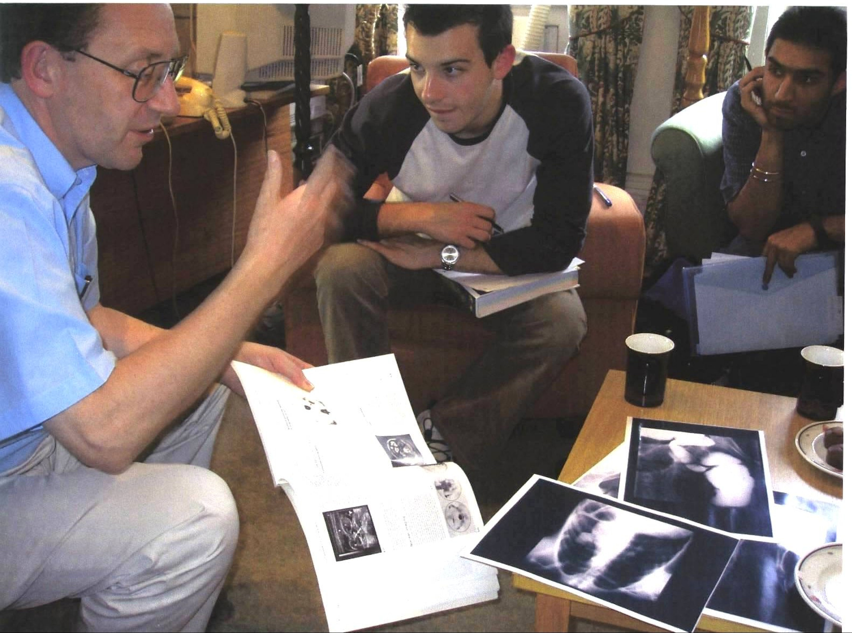
Postgraduate radiology training is high on the agenda in Europe, with a great deal of attention in recent years being given to the harmonisation of educational standards across the continent, but there is a growing feeling within the discipline that radiology should not lose sight of the equally important issue of undergraduate education. Exposing undergraduates to radiology not only serves the obvious and vital purpose of inspiring potential radiologists, but also ensures that students who go on to follow careers in other disciplines are well versed in what radiology can offer and how it operates. In broad terms, the net result is a combination of helping to secure the discipline’s future and making life easier for its practitioners.

However, making sure undergraduates are given sufficient contact with radiology is no easy task. The competing clinical, managerial and academic demands on radiologists’ time and skills, which increase with every year, mean that any additional activities run the risk of being excluded. The time and resources needed, not just to teach, but also to carry out the necessary preparation for effective teaching, can often make it impossible to fit in to an already hectic schedule.

Professor Stephen J. Golding (left) from Oxford will chair today’s Professional Challenges Session on undergraduate teaching.

Professor Stephen J. Golding (left) from Oxford will chair today’s Professional Challenges Session on undergraduate teaching.

Read more…

07
Mar 2013
POSTED BY
POSTED IN ,
DISCUSSION 0 Comments

ECR Today – Cardiac imaging in 2020: reaching new heights

ECR2013_ECRToday_RC303

Watch this session on ECR Live: Thursday, March 7, 16:00–17:30, Room D2

Over the past decade, technological improvements have led to the widespread use of imaging modalities in the prediction, diagnosis and follow-up of coronary disease. Radiologists now have the ability to obtain information on the structure of cardiac muscle with MRI and evaluate cardiac arteries with CT, while hybrid imaging will soon allow them to do both. Cardiac CT will also provide more functional information in the future, and its use will continue to grow. Experts will present the newest and upcoming possibilities of cardiac imaging today at the ECR.

Advances in cardiac CT have brought its use in clinical routine to unprecedented levels. The main reason is that image acquisition optimisation strategies allow radiologists to assess blood vessels with the same efficiency as coronary angiography, non-invasively and almost instantaneously.

ECR_2013_ECRToday_CardiacImaging_1

Read more…

06
Mar 2013
POSTED BY
POSTED IN ,
DISCUSSION 2 Comments

ECR 2013 Focus: the Visibility of the Radiologist

ECR2013_Focus_visibility

With the development of functional imaging, the way patients are imaged has changed, and so has the role of the radiologist. While taking pictures in the basement of a hospital was considered good enough in the twentieth century, radiologists are now required to act as clinicians within multidisciplinary teams. Subspecialisation has become necessary for radiologists in order to keep their leading position in image interpretation. But that is not the only field where radiologists should take the lead, some experts believe. If radiologists were to increase their contribution further up in the diagnostic phase, it would have a significant impact on clinical outcomes and healthcare management. A panel of eminent radiologists will explore ways to do so during a dedicated Professional Challenges Session at ECR 2013.

Professor Any Adam, professor of interventional radiology at the University of London, will speak about the role of the radiologist in the 21st century.

Professor Andy Adam, professor of interventional radiology at the University of London, will speak about the role of the radiologist in the 21st century.

In the past, radiologists were expected to act as technicians with excellent knowledge of the anatomy. But with the creation of functional imaging, things have become more complicated. The capacity to depict and interpret functions using MRI or PET/CT scans calls for more than the ability to read images, and now thorough knowledge of functional processes and organ systems is expected of radiologists.

Meanwhile, many physicians have been increasingly working with images in recent years. For instance surgeons rely on images to prepare for an intervention, and so do radiotherapists to determine the necessary dose and precise location of a tumour to be destroyed. As their experience with reading images is growing, it is likely that these specialists will find it easier to do so without the help of radiologists in the future.

To put it in a nutshell, if radiology is to achieve its full potential, radiologists will have to make a clinical contribution as well. “If radiologists wish to retain their role as experts in image interpretation, they will not only need a thorough understanding of imaging, but also a detailed understanding of anatomy and pathophysiology, and they will need to subspecialise. That is really something we have to take into account in the future. Radiologists will have to get closer to the patient and talk to the referring physician or surgeon. They will have to become more like clinical doctors than they have been until now,” said Andy Adam, professor of interventional radiology at the University of London.

Read more…

ECR 2013 Focus: Imaging in Intensive Care Patients

ECR2013_Focus_carepatients

Intensive care units are special working environments, presenting radiologists with complex cases and patients with severe conditions. Diagnostic imaging examinations and the work of the radiologist have to be adapted towards these special circumstances, which can be one of the biggest challenges when working in an intensive care unit. Today there is a strong need for accurate, clinically relevant radiological input, which often has to be worked out while facing a lack of adequate image material and patients suffering from life-threatening conditions.

Prof. András Palkó from Szeged, Hungary, will chair the session on imaging in intensive care patients.

Prof. András Palkó from Szeged, Hungary, will chair the session on imaging in intensive care patients.

The ECR 2013 Special Focus Session on imaging in intensive care patients, chaired by ESR Past-President, Professor András Palkó from Szeged Medical School in Hungary, will give an up-to-date overview on the use of common imaging methods in the ICU environment. Special Focus Sessions are clearly aimed at in-depth analysis and the promotion of scientific debate between the speakers and their audience.

“The intensive care unit is a very special environment requiring special expertise from both the technicians and the radiologists working in a technically challenging situation. The patients are typically in very severe conditions, frequently unconscious, and almost always connected to life-support and monitoring equipment,” Prof. Palkó pointed out some of the difficulties of working in an ICU.

As a result of this, the majority of imaging examinations are performed on patients with limited ability to cooperate and often at the bedside. Reports are then typically written with insufficient clinical information, based on technically limited images, even though the need for accurate imaging material and radiological information is even greater than in standard clinical settings.

Read more…

ECR 2013 Focus: Personalised Radiology

ECR2013_Focus_PersRad

As acknowledged in last year’s ESR white paper on the subject, the concept of personalised medicine (PM) is becoming an increasingly hot topic. The patient-centred principles of PM have the potential to take over as the dominant philosophy in clinical healthcare in the relatively near future, which would see the focus of the medical world gradually shifting away from the current system of ‘disease care’, towards an approach based on prediction and prevention. However, although most radiologists are aware of the idea of PM and rightly consider medical imaging to already be among the most personalised aspects of healthcare, there is perhaps a need for more awareness of the exact nature of this new paradigm, and specifically a need for recognition – from within and outside the discipline – of the role that medical imaging should play.

ESR President, Prof. Gabriel P. Krestin will chair the Professional Challenges Session on personalised medicine

ESR President Prof. Gabriel P. Krestin will chair the Professional Challenges Session on personalised medicine

A Professional Challenges Session at ECR 2013, chaired by the ESR President, Prof. Gabriel Krestin, will aim to raise awareness of the core principles of PM and highlight the factors that radiologists will need to consider in order to adapt their approach to training, clinical practice and research.

“The whole idea of PM, and the role of imaging within it, is relevant to all of us,” said Krestin, from the Erasmus Medical Centre, Rotterdam, Netherlands. “It is a concept that will gain in importance in the coming years and it will have an increasing influence on the way we work as radiologists. I think many people have heard of PM, but certainly not everyone will have a sound conception of exactly what it is or its full implications for imaging and our daily practice. To be aware of this, and the possibilities that will probably multiply in the coming years, is very important.”

Read more…

ECR 2013 Focus: Justifying CT in Paediatric Radiology

ECR2013_Focus_PaediatricRad

The optimisation and justification of procedures is vital when using CT as an imaging modality; particularly on children, who are more sensitive to ionising radiation than adults. Therefore, it is crucial that all those who use CT understand the physics behind the equipment and ultimately use this understanding to minimise the potential risks while maximising the potential benefits to each individual patient. Patients should also be informed of the risks and benefits of undergoing a CT scan. World-renowned experts will explain these issues in detail during a Special Focus Session at ECR 2013.

“Not all radiologists and technicians are aware of the latest dose reduction strategies. Some are not necessarily so well-informed and perhaps do not realise how important this is. We believe that it is a question of trying to get everybody to a certain level of knowledge and expertise,” said Dr. Catherine Owens, paediatric radiologist and CT unit lead at Great Ormond Street Children’s Hospital in London, U.K.

Paediatric MDCT  Chest Imaging Techniques

Fig. 1

Read more…

ECR 2013 Focus: Organs From A to Z: Heart

ECR2013_Focus_8_AZHeart

Heart disease affects a very large number of people worldwide, and the consequences can be serious and even lethal. Here, and perhaps more than in many other areas of medicine, imaging has helped to improve treatment and prevention. It does so by detecting the disease at an early stage, sometimes even before its emergence, especially in patients at risk of ischaemic heart disease.

Today, diagnosing cardiac patients has become routine for many radiologists. However, some of them may not know of recent developments in this field and they may need to refresh their knowledge. A panel of experts will update both general and specialised radiologists with the latest information available on cardiac imaging, during the dedicated Mini Course ‘Organs from A to Z: Heart’ at ECR 2013. After an introduction to heart anatomy and the main imaging protocols, the course will focus on valvular diseases and cardiomyopathies; two pathologies commonly encountered in radiology practices.

Figure 1

Figure 1:
A) Example of a dilated cardiomyopathy (DCM). Cine-MR images in four-chamber view (left) and short-axis view (right) at end-diastole show significant dilatation of the LV cavity. Ejection fraction was <35% in this patient. (RA = right atrium; LA = left atrium; RV = right ventricle; LV = left ventricle)
B) Example of an asymmetrical, apical hypertrophic cardiomyopathy (HCM). Cine-MR images in a four-chamber (left) and two-chamber view (right) in systole show a markedly thickened left ventricular myocardium predominantly of the apex, as compared with the basal segments (RV = right ventricle; LV = left ventricle).

Read more…

ECR 2013 Focus: Imaging of the Mind

ECR2013_Focus_7_ImagingMind

The brain remains undoubtedly one of the most mysterious organs of the human body. Magnetic resonance imaging has helped to unveil some of its secrets, and major advances have been made in understanding how the brain functions. Recent developments with resting fMRI (rfMRI) and diffusion MRI (dMRI) indicate that scientists are beginning to see beyond the brain: they have actually started to visualise the human mind. This new information is particularly relevant for understanding complex processes such as dementia, autism and depression. It is also proving increasingly central to the diagnosis of comas and chronic disorders of consciousness.

Leading researchers will discuss where the latest advances have led them and what the future will bring in a dedicated New Horizons Session during ECR 2013. FMRI has been used for over twenty years to visualise changes in brain activity by comparing a task versus a control task, and showing and quantifying how much brain activity is involved in the process. The recent addition of rfMRI enables researchers to track networks that are randomly active. A patient lying in a scanner with no particular task to perform will usually start thinking about the trivialities of the day and go from one thought to the other (“Did I close the door before I left? What am I doing here?” etc.). Neuroresearchers can track this mind mumbling with complex mathematics and extract information from what they call the default mode network.

Fig. 1: Differences in functional connectivity from rfMRI between autistic patients and age- and gender matched controls: the major disconnection is between the cerebellum and frontal language areas.

Fig. 1: Differences in functional connectivity from rfMRI between autistic patients and age- and gender matched controls: the major disconnection is between the cerebellum and frontal language areas.

Read more…

ECR 2013 Focus: Sessions for Radiographers

ECR2013_Focus_6_Radiographers

The quality of the ECR’s sessions for radiographers has been given a welcome seal of approval from the European Federation of Radiographer Societies (EFRS) who recently elected the ECR as their official annual scientific meeting. EFRS president, Prof. Graciano Paulo, from the college of health technology of Coimbra, Portugal, has been coming to the ECR for more than a decade and firmly believes the upcoming congress boasts one of the best selections yet for radiographers. Here he gives his personal preview of ECR 2013 and each of these sessions, all of which you can find in the ECR 2013 Interactive Programme Planner by searching for ‘radiographers’.

Read on for Prof. Paulo’s preview of all the sessions for radiographers at ECR 2013 …

Read more…

ECR 2013 Focus: ESR Meets the E-AHPBA

ECR2013_Focus_Final5_EAHPBA

The ‘ESR Meets’ programme, as well as being an extremely popular part of every ECR, serves a very valuable purpose. The sessions have not only reinforced relationships with numerous European national societies since the programme’s introduction in 2003, but they have also represented the ESR’s first official connections with several societies, sometimes from other continents and, more recently, from other disciplines. This year’s partner society, the European-African Hepato-Pancreato-Biliary Association (E-AHPBA) takes the ‘ESR Meets’ programme one step further …

E-HPBA president, Prof. Pierre-Alain Clavien, from Zurich, Switzerland

E-HPBA president, Prof. Pierre-Alain Clavien, from Zurich, Switzerland

Read more…

07
Feb 2013
POSTED BY
POSTED IN ,
DISCUSSION 0 Comments