Incoming ESR President lays out clear vision for ECR 2020

By Julia Patuzzi

It is a well-established tradition that on the final day of the congress, ECR Today looks ahead to next year’s ECR. We therefore spoke with Professor Boris Brkljačić from Zagreb, Croatia, the incoming ESR President, who is in charge of ECR 2020. He shared with us some of his ideas and plans for the next European Congress of Radiology.

ECR Today: Professor Brkljačić, the first visual impression of the next congress is always the congress poster. For ECR 2020 you chose artwork by the award-winning Canadian illustrator Peter Diamond, depicting a young woman looking at a small object floating just above her cupped hand. Can you tell us a little about how this particular design came about?

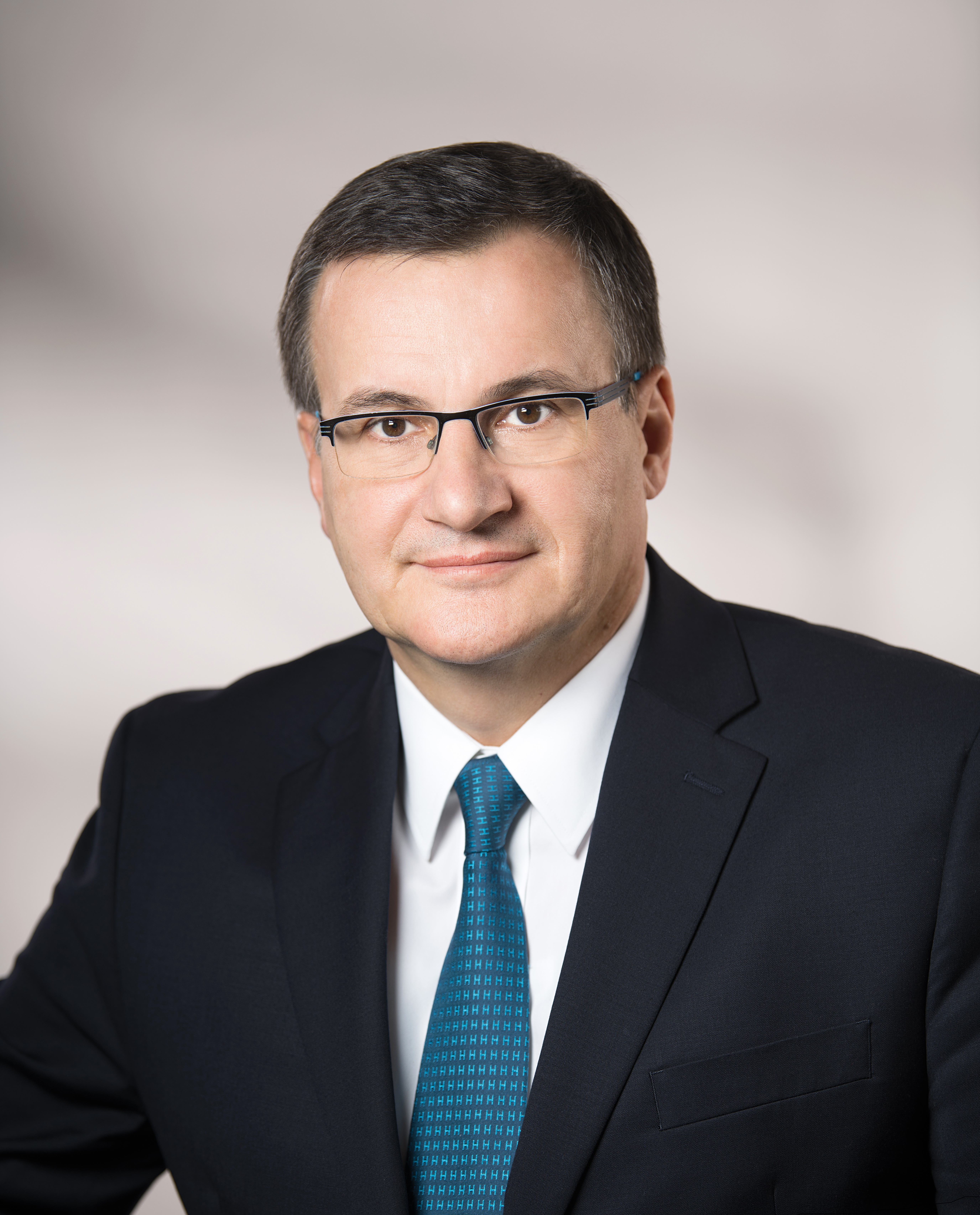
Incoming ESR President Boris Brkljačić is professor of radiology and vice-dean at University of Zagreb School of Medicine, Zagreb, Croatia, and chair of the Department of Diagnostic and Interventional Radiology of University Hospital ‘Dubrava’ in Zagreb.

Boris Brkljačić: The ESR Office provides several options for the congress poster, created by professional designers, and the Congress President and PPC members select one. The selected solution was the best among the proposed options. It resembles Rembrandt’s artwork, with sharp light and dark contrast, and is in good accordance with the slogan for ECR 2020: ‘A Clear Vision for Radiology’. The small floating object represents artificial intelligence, which will be an important topic at the congress, and the names of the ECR 2020 ‘ESR meets’ countries are visible at the bottom of the poster. The 2020 poster contains fewer colours and illustrations compared to the 2019 poster, and is concordant with the visual style of the ESR’s main scientific publication, the journal European Radiology.

ECRT: As the new ESR President, you are also chairperson of the Programme Planning Committee for ECR 2020, which has already been working on preparing the scientific programme for a few months. Can you tell us something about the highlights of the 2020 programme or any specific focus we can expect?

BB: I am very fortunate to have selected excellent Programme Planning Committee members, who are hard-working and dedicated experts in their fields. Planning has already been running at full speed for a few months in order to create a well-balanced programme of very high-quality professional, educational and scientific content. New Horizons Sessions, State of the Art Symposia and Special Focus Sessions have already been selected and mostly created; they are very relevant and balanced, so that young radiologists and experts in particular radiological fields will have interesting sessions to choose from in all areas of radiology. Emerging and hot topics will be covered, like lung cancer screening, artificial intelligence, stroke diagnosis and treatment, and many others. I expect that the plenary/honorary sessions should be the highlight of the congress, as they are interesting for all participants, regardless of their age and expertise. Read more…

Major interventional radiologist receives ESR Gold Medal

Anna-Maria Belli, Professor of Interventional Radiology at St. George’s Hospital in London, UK, will be presented with the Gold Medal of the European Society of Radiology today. Ahead of the ceremony, she shared her views on which directions interventional radiology should take.

Interventional radiology (IR) procedures initially broke into the field of peripheral arterial disease by opening up blocked arteries and establishing angioplasty as a valid, alternative, minimally invasive therapy for those either unfit or unsuitable for standard bypass surgery. It has now become an accepted therapy, replacing open surgery in many situations and responsible for saving limbs from amputation.

IR specialist Prof. Anna-Maria Belli from London will receive the ESR Gold Medal during the ECR 2019 Grand Opening today at 17:45 in Room A.

As experience and skill with arterial catheterisation advanced, so did arterial embolisation, which is used with the opposite intention from angioplasty by selectively occluding arteries. Initially this was an emergency procedure used to treat life-threatening haemorrhage, making it difficult to train in. However, with its expanded indication in the treatment of vascular tumours, it has become a common elective procedure and it has been one of Belli’s special areas of research during the four decades in which she has practised as an interventional radiologist. “Embolisation is now an alternative treatment which may replace standard surgical options, e.g. in the treatment of fibroids and benign prostatic hyperplasia,” she said.


Non-vascular interventional radiology has also grown in leaps and bounds, particularly in the management of cancer with interventional oncology. “The new technologies being introduced into interventional oncology are amongst the most exciting developments and indicate a very strong future for IR,” she believes. Read more…

ECR continues to think outside the box and adds the Cube

For young physicians, interventional radiology (IR) may seem as enigmatic as a Rubik’s cube. But the cube can be solved with the right algorithm, according to Dr. Maximilian de Bucourt, head of angiography at Charité Campus Benjamin Franklin in Berlin.

“What we do in IR is exactly like a Rubik’s cube: you follow the algorithm and solve the problem. In IR, if you do the procedure steps over and over again and use the rules, most of the time you can get the solution for the patient,” he said.

Dr. Maximilian de Bucourt is head of angiography at Charité Campus Benjamin Franklin in Berlin, and one of the specialists behind the Cube.

Together with Prof. Christian Loewe from Vienna, de Bucourt imagined ‘the Cube’, a workshop aimed at introducing young physicians to the tools and techniques used in IR. With its focus on hands-on activities, including simulated procedures and interactive demonstrations, the Cube is fulfilling its goal of acquainting residents with this unique sub-specialty.

“The Cube is for young people who are thinking of becoming IR specialists, but find it too long before they can deploy a stent or manoeuver a catheter inside an artery. After medical school, basic radiology training and dedicated IR training with a teacher willing to let them perform major interventions, they still need to learn how to manage complications. The Cube is all about expediting this process, by enabling students and residents to get their hands on the products earlier,” he explained. Read more…

Image-guided interventions: a key pillar in cancer care

Watch this session on ECR Live: Wednesday, March 1, 16:00–17:30, Room F2
#ECR2017

 There is hardly any area of hospital medicine where interventional radiology (IR) has not had some impact on patient management. The range of conditions that can be treated using interventional radiology techniques is continually expanding.

In today’s session, experts will provide an insight into image-guided interventions in oncology with a particular focus on illustrating the importance of quality assurance in image-guided oncological interventions and their effect on treatment outcomes.

In recent years, IR has played a vital role in the field of oncology, and alongside medical, surgical and radiation oncology it constitutes a key pillar in cancer care. Vascular and non-vascular procedures such as transarterial chemoembolisation, radiofrequency ablation (RFA), microwave ablation, radioembolisation, cryoablation and high-intensity focused ultrasound (HIFU) are delivered locally, minimise damage to nearby tissue and avoid the systemic side effects of chemotherapy.

Colorectal lung metastasis before CT – guided microwave ablation.

For the interventional radiologist providing oncologic therapies it is essential to understand the rapidly changing field of oncology and to have a broad knowledge of oncologic diseases and available therapies to treat them. Radiologists providing image-guided interventions in oncology have an outstanding understanding of imaging as well as a diversity of interventional skills. However, they lack formal training in oncology and an understanding of chemotherapy and radiotherapy, according to Prof. Andy Adam from the Department of Radiology at Guy’s and St. Thomas’ Hospital in London. Read more…

ECR 2013 Rec: Detection of HCC and liver metastases with BR14: final results of a multicentre phase IIA study #SS201b #B0232

ECR2013Rec_Blog_Cover_B0232_SS201b

B-0232 Detection of HCC and liver metastases with BR14: final results of a multicentre phase IIA study

J. Hohmann, A. Müller, J. Skrok, K.-J. Wolf, A. Martegani, C.F. Dietrich, T. Albrecht | Thursday, March 7, 14:00 – 15:30 / Room I/K

Purpose: The study was primarily designed to find the optimal dose range of BR14 to detect malignant focal liver lesions. Secondary objectives were the evaluation of the safety profile and comparison with contrast-enhanced MRI (CE MRI).
Methods and Materials: 25 patients (9f, 16m, mean age: 66y) with known HCC or liver metastases were examined in three centres during a time period of three months. Each patient underwent a baseline and at least three contrast-enhanced US (CEUS) with ascending dose levels (0.25ml, 1.0ml, 4.0ml) of BR14. CE MRI was done 4 weeks prior or post-study examination. Lesions were recorded in on a liver map, with respect to localisation, size and suggested lesion type. Examination quality was documented and safety parameters were assessed.
Results: The number of lesions detected with BR14 CEUS increased with dose, while the number of missed lesions and the lesion size decreased. Despite the increasing contrast enhancement no other image quality parameter showed a substantial difference. No significant changes were found for the analysed safety parameters and no serious adverse events were reported.
Conclusion: We finally conclude that the recommended dose level of BR14 is between 1.0 ml and 4.0 ml for which the lesion detection was comparable to the CE MRI. In addition, we found a higher number of especially small lesions with higher doses of BR14 which might be due to a higher sensitivity of CEUS for the detection of liver metastases. However, this is an interesting and debatable finding.

ECR 2013 Rec: A. RF ablation #RC709 #A187

ECR2013Rec_Blog_Cover_A187_RC709

A-187 A. RF ablation

 J. del Cura | Friday, March 8, 16:00 – 17:30 / Room N/O

RF ablation is currently indicated in HCC as curative treatment in Child-Pugh A-B patients with: single <2 cm nodule and not candidates for transplation, 1-3 nodules <3 cm and not candidates for resection or transplantation. RF can be also performed in patients waiting for liver transplantation. Some studies suggest that survival does not differ between RF ablation and resection in 5 cm because of the high possibility of recurrence. Different types of electrodes can be used: internally cooled, cluster, expandable, with saline instillation. Although results can be good with any of them, every type of device requires a different technique of ablation. Obtaining a margin of at least 0.5 cm of ablated tissue around the tumour is key to avoid recurrences. Combined treatments like combining chemoembolization or PEI with RFA can be useful to increase the ablation volume. Published data show a pooled 5-year survival of 48-55%, with better outcomes in Child-Pugh A patients. In candidates for surgery, 5-year survival is similar to resection: 76 %. RFA is safe: major complications appear in 10 % and reported mortality is 0.15%. Tumours located subcapsular or near major vessels, biliary tree or bowel are more prone to complications. Laparoscopic ablation can be an alternative in these cases. Imaging follow‑up with CT, MRI or CEUS is performed to assess the outcome and detect recurrences, new lesions or complications. Although not well established, most protocols include an immediate post-procedure imaging, 1-month follow-up and explorations every 3 or 6 months for 2-3 years.

ECR 2013 Rec: Biliary procedures #SF14a #A445

ECR2013Rec_Blog_Cover_A445_sf14a

A-445 Biliary procedures

M. Krokidis, A.A. Hatzidakis | Sunday, March 10, 14:00 – 15:30 / Room F1

Palliative Percutaneous Transhepatic Biliary Drainage (PTBD) is a therapeutic procedure leading to drainage of the obstructed bile duct system. If endoscopy is not possible and if patient is inoperable, then the percutaneous treatment is indicated. Drainage of the bile ducts is performed with a small plastic multiple hole pigtail catheter. Self-locking catheters are preferred in order to minimize the dislocation risk. The percutaneous catheter is pushed through the malignant stricture, so that bile is draining through the catheter towards the bowel loops. Technical success rate of percutaneous biliary drainage can reach nearly 100 % in experienced hands, while the major complications rate is usually lower than 5 %. Clinical efficacy is usually lower, but still over 90 %. The drainage procedure can be extended with the placement of a permanent metallic stent, which keeps the stenosed biliary duct patent, without need for a catheter. Metallic biliary stents have been proved as the best palliative treatment of non-resectable malignant obstructive jaundice, allowing longer patency rates than plastic endoprostheses. The technique is safe, with low-complication rate and procedure-related mortality between 0.8 and 3.4%. Still controversial remains in the timing between initial drainage and metallic stent placement, as well as the question of balloon dilatation before stent insertion. There is evidence that if the initial transhepatic drainage is completed without causing any severe complications, especially bleeding in form of haemobilia, primary metallic stenting can follow as a single-step procedure.


ECR 2013 Rec: Chairman’s introduction #A339 #RC1109

ECR2013Rec_Blog_Cover_RC1109_A339

A-339 Chairman’s introduction

M. Krokidis, A.A. Hatzidakis | Saturday, March 9, 16:00 – 17:30 / Room N/O

There are some basic IR techniques everybody needs to know before starting to treat biliary diseases. Τhese are Percutaneous Transhepatic Cholangiography (PTC) and Percutaneous Transhepatic Biliary Drainage (PTBD). These basic procedures are essential first for opacification and then for getting access into the biliary tree. After accessing the biliary system, decisions must be made about the need for further interventional procedures. Presence of benign or malignant strictures, stones or other kind of disease are leading us to the necessary actions, such as dilatation, stenting or lithotripsy. Dilatation of stenosed parts of the biliary system is performed after negotiation of the stricture or occlusion with help of high-pressure angioplasty balloons of variant sizes. Balloons of 8-14 mm width can be used alone or two parallel to each other. Stenting is rarely needed in benign biliary disease and usually in cases where multiple dilatations are not responding. Percutaneous lithotripsy with or without cholangioscopic assistance is a wide-used technique for clearance of biliary stones or fragments. Extraction balloons or baskets and special lithotripsy devices are commonly used for impacted or large calculi.

ECR 2013 Rec: Hepatic parenchymal and vascular contrast improvement in super-delayed phase images of Gd-EOB-DTPA-enhanced MRI #B0984 #SS1801b

ECR2013Rec_Blog_Cover_SS1801b_B0984

B-0984 Hepatic parenchymal and vascular contrast improvement in super-delayed phase images of Gd-EOB-DTPA-enhanced MRI

S. Kobayashi, O. Matsui, T. Gabata, W. Koda, T. Minami, K. Kozaka, A. Kitao | Monday, March 11, 14:00 – 15:30 / Room I/K

Purpose: To elucidate the parenchymal and vascular contrast improvement effect of super-delayed phase (SDP) images of Gd-EOB-DTPA (EOB)-enhanced MRI in poor hepatobiliary phase (HBP) image cases special focus on Child-Pugh (CP) classification.
Methods and Materials: 76 cases, who have examined EOB-enhanced MRI for closer examination of hepatic lesions, and taken SDP images approximately 90 minutes after iv administration of EOB because of poor HBP image are subjected to this study. 20 hepatobiliary disease cases who had also taken SDP images which show normal HBP images were used as control. Hepatic vascular/parenchymal enhancement ratios (ER) were defined as signal intensity (SI) of intrahepatic vessel / SI of liver. ER of HBP and SDP were calculated and compared between each CP class liver damage groups. Chi square test was used for statistics and p<0.05 was considered statistical significant.
Results: In poor HBP cases (n=76), ER of HBP and SDP were 0.88±0.16 and 0.64±0.16. In control cases (n=20), ER of HBP and SDP were 0.54±0.08 and 0.39±0.06. ER of HBP and SDP in CP-A poor HBP (n=27), CP-B poor HBP (n=47), CP-C poor HBP (n=2) were 0.83±0.14 and 0.60±0.13, 0.90±0.16 and 0.65±0.16, 1.03±0.16 and 0.99±0.19, respectively (all combinations except CP-C showed significance difference).
Conclusion: In most of the poor HBP image cases, SDP image improve parenchymal and vascular contrast except CP-C liver damage cases.

ECR 2013 Rec: Pre-therapeutic radiological evaluation #A245 #RC809

ECR2013Rec_Blog_Cover_RC809_A245

A-245 Pre-therapeutic radiological evaluation

J. Raupach, O. Renc, P. Hoffmann, J. Zizka; | Saturday, March 9, 08:30 – 10:00 / Room N/O

Endovascular abdominal aortic aneurysm repair (EVAR) was introduced over 20 years ago to primarily treat old and sick patients. Due to technical improvements and satisfactory clinical results of this technology, the number of patients treated with stent-grafts is steadily increasing. There is also tendency to use this therapy for ruptured abdominal aortic aneurysms. Pre-operative assessment of aortic morphology regarding suitability for stent-graft implantation is, therefore, an important challenge for every radiologist now. Main limitation of EVAR is unfavourable anatomy of landing zones and access vessels. Gold standard for EVAR planning is contrast-enhanced CTA. Alternative modality for patients with contraindications for CT, such as renal impairment, is unenhanced MR with steady-state free precession sequence. A number of 2D or 3D reconstructions are generated to provide information about the aneurysm morphology. Dedicated vessel analysis and planning software can be applied. Usually, axial images and thin MPR reconstructions are sufficient in emergent cases. Proper stent selection is a domain of operator and is still matter of his/her experience. The planning procedure can be subdivided into 4 different sections: infrarenal neck, aneurysmal sac, aortic bifurcation and access vessels. There are several critical and rules which must be obeyed during the evaluation process and general radiologists should be aware of. The presentation will review main inclusion and exclusion criteria for EVAR.