
Dear Friends,
To finish the chapter on male breast disease I am showing mammograms with selected pathology in four different males with palpable retro-areolar lesions. Try to match each case (A-D) with the correct diagnosis.
1. Gynecomastia
2. Carcinoma
3. Epidermoid cyst
4. Simple cyst
Read more…

Dear Friends,
This week I’m showing a new ‘Face the Examiner’ case. Presenting the radiographs of a 49-year-old woman with dyspnea.

Dear Friends,
This week we have the case of a 45-year-old man, who is an alcoholic with abdominal pain, jaundice, and weight loss.
Possible diagnoses:
1. Duodenal neoplasm
2. Focal pancreatitis of pancreatic head
3. Pancreatic neoplasm
4. None of the above
Read more…

Dear Friends,
Today I’m showing a case of a 63-year-old man with left heart dysfunction and angor.
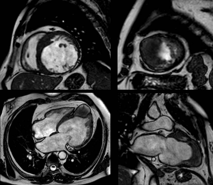
Read more…

Dear Friends,
This week I’m presenting the case of a 9-year-old child with pain in the leg after trauma.
Diagnosis:
1. Aneurysmal bone cyst
2. Simple bone cyst
3. Giant-cell tumour
4. Osteosarcoma
Read more…

Dear friends,
This week I’m presenting you another new ‘Face the Examiner’ case which simulates a real examination. Showing radiographs of a 35-year-old male with high fever and left pleuritic pain.

Dear Friends,
This week I’m showing the case of a 26-year-old male presenting with chest pain.
Read more…

Dear Friends,
As some of you may be taking the Diploma examination during ECR 2013, I’d like to introduce a new type of post that I call ‘Face the Examiner’. Its purpose is to simulate a real examination: images will be shown and you will be asked to describe the findings. You should then offer a differential diagnosis and suggest a procedure that will confirm your preferred option.
The only difference with respect to a real examination is that you will be given the correct answers during the exercise. I’ll try to keep it simple by not giving long lists of possible diagnoses and by making sure the possibilities are coherent with the imaging features.
Our first ‘Face the Examiner’ case concerns preoperative chest radiographs in a 75-year-old man with prostate carcinoma.

Dear Friends,
This week, we have an oncologic patient with liver nodule detected on US examination. Below are the images from an MRI examination.
Possible diagnoses:
1. Liver hemangioma
2. Hepatocellular carcinoma (HCC)
3. Liver metastasis
4. Focal nodular hyperplasia (FNH)
Read more…

Dear Friends,
Showing chest radiographs of an 81-year-old male with multiple bone fractures after a car accident. There is a rounded well-defined opacity in the posterior costophrenic sulcus. What do think it is?
1. Carcinoma of the lung
2. Bochladek’s hernia
3. Diaphragmatic cyst
4. All of the above
Read more…