Education central to improving imaging data quality in oncology clinical trials

Watch this session on ECR Live: Thursday, March 2, 16:00–17:30, Room X
#ECR2017

Imaging data is key in multicentre clinical trials for cancer research but quality control is currently a major impediment, bringing the validity of the trials into question and potentially impacting on the quality of drugs put on the market, a panel of experts will argue today in a session held by the ESR and the European Organisation for Research and Treatment of Cancer (EORTC) at the ECR.

Imaging is increasingly contributing to cancer research thanks to the development of innovative techniques that depict functional and molecular processes. In most oncological clinical trials, imaging is now the primary criteria used to evaluate progression of disease or efficiency of the drug being tested.

The best way to obtain valuable imaging measurements is to involve the imagers who take part in these trials and educate the clinician investigators, experts will explain in the session.

When it comes to imaging in cancer research, a number of issues take centre stage. Difficulties associated with integrating imaging biomarkers into trials have been neglected compared with those relating to the inclusion of tissue and blood biomarkers, largely because of the complexity of imaging technologies, safety issues related to new contrast media, standardisation of image acquisition across multivendor platforms and various post-processing options available with advanced software, as reported recently in The Lancet by the EORTC and leading researchers.

Read more…

01
Mar 2017
POSTED BY
POSTED IN ,
DISCUSSION 0 Comments

Eminent neuroradiologist to give honorary lecture at ECR 2017

Watch this session on ECR Live: Thursday, March 2, 12:15–12:45, Room A
#ECR2017

In recognition of his significant impact on the field of neuroimaging and his service to organised radiology, Professor Mauricio Castillo from Chapel Hill, NC, United States, has been invited to deliver the Wilhelm Conrad Röntgen Honorary Lecture ‘Dissatisfaction, burnout and inequality: three major challenges in radiology’ at ECR 2017.

Professor Mauricio Castillo from Chapel Hill, United States

Professor Mauricio Castillo from Chapel Hill, United States, will deliver the Wilhelm Conrad Röntgen Honorary Lecture today at 12:15 in Room A.

Mauricio Castillo is the James H. Scatliff distinguished professor of radiology, chief and programme director of neuroradiology at the University of North Carolina in Chapel Hill. He is currently president of the American Roentgen Ray Society.

Originally from Guatemala, Prof. Castillo completed his radiology and neuroradiology training at the University of Miami School of Medicine, Jackson Memorial Medical Center, and Emory University School of Medicine, Affiliated Hospitals in Atlanta respectively.

His specialty and subspecialty choice was in large part motivated by his mentors.

“When I was a medical student, I was fortunate to work in the only teaching hospital that had a CT scanner and two board-certified radiologists. I was very impressed by the impact of imaging in patient care and by the way these two professionals used their large base of knowledge of medicine to generate a differential diagnosis based on imaging findings. Later on, while I was a resident, I developed a close relationship with Dr. Robert Quencer, which led me to become a neuroradiologist. One should never underestimate the influence we may have on our trainees and the influence our mentors have had on us,” he said.

Prof. Castillo’s research interests include paediatric neuroimaging, application of new imaging techniques and medical literature editing. Read more…

Prominent Brazilian radiologist to shed light on Zika virus imaging at ECR 2017

In recognition of her major achievements in neuroimaging and advancement of the field, Professor Maria de Fátima Viana Vasco Aragão from Recife, Pernambuco, Brazil, will present the Guest Lecture ‘Breaking News from Latin America: How to recognise Zika virus infections on imaging studies’ at ECR 2017.

Maria de Fátima Viana Vasco Aragão is professor of radiology at the Maurício de Nassau University and Scientific Director of the Multimagem Diagnostic Centre in Recife, Brazil. She is also financial director of the Diagnostika Endoscopy and Radiology Clinic in Recife.

Prof. Vasco Aragão received her medical degree from Pernambuco Federal University in 1987 and completed her residency in radiology at Ribeirão Preto Faculty of Medicine, University of São Paulo. She did fellowships in CT and MRI and in neuroradiology at the Med Imagem Beneficencia Portuguesa, São Paulo, and later completed a research fellowship at Mount Sinai Hospital Center in New York, US.

She always knew she would become a doctor and chose radiology because she recognised early on the field’s value in diagnosis and treatment.

Maria de Fátima Viana Vasco Aragão is professor of radiology at the Maurício de Nassau University and Scientific Director of the Multimagem Diagnostic Centre in Recife, Brazil.

Maria de Fátima Viana Vasco Aragão is professor of radiology at the Maurício de Nassau University and Scientific Director of the Multimagem Diagnostic Centre in Recife, Brazil.

“My mother is a retired biologist and biology teacher. I have never forgotten the moment she taught me what a cell was. In my family, educators predominated and my great aunt was the first woman in Pernambuco, my home state, to graduate from medical school, in 1935. Perhaps on account of this family background I made up my mind to become a doctor. As an intern student of medicine, I was very impressed when I took my patients to be examined by CT and US. Even though there was only one CT scanner in my city, I succeeded in having all my intern patients examined, when indicated. My tutors were impressed by the swiftness with which cases were resolved. Thus I learned that radiology not only helps to quickly define diagnosis in most patients, but also provides a clear prognosis for the most adequate treatment. At the end of the course I began considering the possibility of specialising in radiology, even though 29 years ago, for most people, a radiologist was not a doctor. Shortly after graduation, I sat for a test and won a place in radiology at the first attempt, and radiology has been my passion ever since. I would like to say that I am grateful for having had great mentors, including the neuroradiologist Dr. Sérgio Santos Lima, who was my director during my fellowship in the Med Imagem Beneficencia Portuguesa, and the head and neck radiologist, Dr. Peter Som, who was one of my directors during my research fellowship at Mount Sinai Hospital Center New York,” she said.

Read more…

ECR on Demand Preview: Thoracic emergencies #E³ 1520 #A-485

ECROnDemandPreview_Blog_Picture_06

E³ 1520 – Thoracic emergencies, A-458 B – Pulmonary

A short preview of lecture A-458 ‘B. Pulmonary’, from the session E³ 1520 ‘Thoracic emergencies’ at ECR 2014, given by C.M. Schaefer-Prokop from Amersfoort, Netherlands. Watch the whole lecture and many more at http://ipp.myESR.org Direct link: http://bit.ly/Thoracic_emergencies

Sunday, March 9, 16:00 – 17:30 / Room A

Abstract: Acute respiratory failure can have multiple underlying causes including infection, fluid overload, immunological diseases or exacerbation of preexisting lung disease. Since the clinical symptoms are nonspecific, imaging plays an important role. The first imaging method is mostly the chest radiograph, easy to access and to obtain, but non-diagnostic in many cases. (HR)CT offers more possibilities to define the differential diagnosis. The option of this interactive workshop will be to get familiar with the spectrum of diseases that can cause acute respiratory failure and learn about key findings in radiography as well as CT to reduce the differential diagnosis. The interaction between preexisting lung disease, clinical information (e.g. chemotherapy, rheumatoid arthritis, COPD) and imaging findings will be discussed using clinical case studies. Options and also limitations of imaging findings will be illustrated. The following scenarios will be taken into account: acute cardiac failure and various appearances of oedema; acute immunological-toxic disorders including drug-induced lung disease and inhalational injuries; exacerbations of preexisting lung disease including fibrotic and obstructive lung disorders; severe infections causing respiratory failure and their complications.

28
Oct 2014
POSTED BY
POSTED IN ,
DISCUSSION 0 Comments

ECR on Demand Preview: The treated spine and joints #E³ 920a #A-246

ECROnDemandPreview_Blog_Picture_02

E³ 920a – The treated spine and joints, A-246 A. Imaging of the postoperative spine (P.N.M. Tyrrell)

A short preview of lecture E³ 920a’ The treated spine and joints’, from the session A-246 ‘A. Imaging of the postoperative spine’ at ECR 2014, given by P.N.M. Tyrrell from Oswestry, United Kingdom.

Watch the whole lecture and many more at http://ipp.myESR.org
Direct link: http://bit.ly/The_treated_spine_and_joints


Saturday, March 8, 10:30 – 12:00 / Room A

Abstract:

Spinal surgery is most frequently performed to decompress (disc herniation, stenosis, malignant infiltration), fuse and stabilise (particularly following trauma or infiltrative destructive processes) and correct deformity. Often, there may be a combination of these procedures at one operation. Surgical instrumentation or bone graft is sometimes employed. Patients may present themselves with symptoms early or late following the procedure. This interactive session seeks to address the variety of surgical procedures undertaken and subsequently imaged post-operatively because of symptoms. The session aims to help one to understand and become familiar with the expected post-operative imaging appearances related to the surgical procedure, learn about abnormal pathological features as a cause of symptoms in the acute and more chronic situation and explore the diagnosis and differential diagnosis. This may include post-operative fibrosis versus recurrent disc herniation versus post-operative infection. Failure of fusion due to failure of instrumentation or inadequate take of bone graft can give rise to pseudoarthrosis. Recurrent stenotic symptoms may relate to an inadequate decompression, recurrent disc herniation, post-operative haematoma, extension of a malignant process or ischaemic damage.

Advances in forensic imaging bring new opportunities for radiology

ECR2013_ECRToday_Sunday_Cover2

Watch this session on ECR Live: Sunday, March 10, 14:00–15:30, Room Z

The ability to spot pathological imaging findings among normal post-mortem signs of degeneration may not be a universal skill among radiologists, but it could prove to be a useful, if not essential one. As the reliability of modern forensic imaging rapidly improves, forensic pathologists are increasingly seeking the help of radiologists to examine bodies non-invasively, so they should be prepared to answer that call, according to experts who will speak in a Special Focus Session on ‘Advances in forensic imaging’ today. The development of techniques such as spiral volumetric CT and, more recently, MRI, have dramatically improved the ability of radiologists to determine causes of death and detect other crucial post-mortem signs, providing an invaluable service that can supplement, and in some cases replace, traditional autopsy.

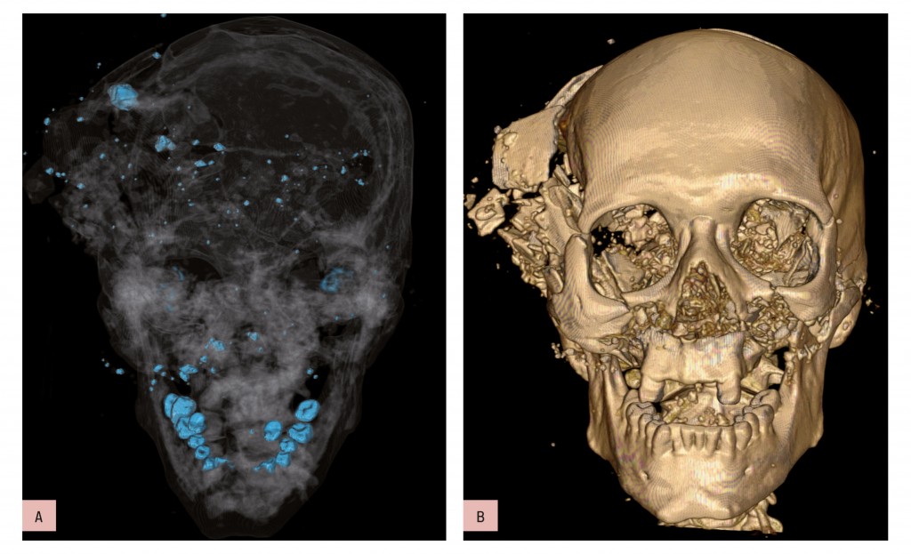
Bone and metal maximum intensity projection (MIP) reconstruction (a) and volume-rendered (VR) 3D-CT reconstruction (b). Homicidal death: characteristic bony and metallic fragments on the exit side of the skull, where the bullet caused a large loss of brain, leading to shattering of the skull.

Bone and metal maximum intensity projection (MIP) reconstruction (a) and volume-rendered (VR) 3D-CT reconstruction (b). Homicidal death: characteristic bony and metallic fragments on the exit side of the skull, where the bullet caused a large loss of brain, leading to shattering of the skull.

Read more…

10
Mar 2013
POSTED BY
POSTED IN ,
DISCUSSION 2 Comments