A-093 Cardiomyopathies
P. Sipola | Friday, March 8, 08:30 – 10:00 / Room L/M
Cardiac magnetic resonance imaging (CMRI) is highly valuable in the differential diagnosis of cardiomyopathies. MRI diagnosis is based on cine imaging of cardiac function, T2-weighted imaging of oedema and late gadolinium-enhanced (LGE) patterns of scar tissue. Hypertrophic cardiomyopathy (HCM). Left ventricular hypertrophy (LVH) is typically located in basal septum and anterior wall but has variable expression (diffuse, localized). Associated abnormalities include left ventricular (LV) high-ejection fraction (EF), mitral valve abnormalities, apical aneurysm, and right ventricular (RV) hypertrophy. Scattered intramyocardial LGE may occur in various patterns. The differential diagnoses in patient with hypertrophic phenotype include pressure overload hypertrophy, amyloidosis, sarcoidosis, and Fabry’s disease. LGE patterns is useful in differentiation. Dilated cardiomyopathy (DCM): Dilated LV end-systolic volume and impaired EF% are characteristics. Non-ischaemic DCM typically shows no LGE (in contrast to ischaemic cardiomyopthy). Sometimes faint midwall enhancement can be observed, which has prognostic value. Presence of extensive non-compacted myocardium indicates non-compaction cardiomyopathy. Arrhythmogenic right ventricular cardiomyopathy (ARVC): The RV volume is enlargened and akinetic RV segments can be seen. Local bulging or dyskinesia in conjunction with fatty infiltration and LGE is typical. Restricted cardiomyopathy (RCM): Enlargened atrias and normal sized ventricles with preserved EF% and no LGE are characteristics. Myocarditis: LV systolic function is typically lowered but may be normal. T2 images may show increased signal. LGE limited to the subepicardial myocardium is highly suggestive of myocarditis. Iron overload cardiomyopathy: Cine imaging is used to assess LV global function and T2*-weighted imaging to quantitate ventricular iron deposition.
Watch this session on ECR Live: Saturday, March 9, 16:00–17:30, Room G/H
Advanced MR imaging techniques such as perfusion and functional imaging have been a great help in improving the diagnosis and staging of brain tumours. Unlike conventional MR techniques, advanced MR techniques can be used to obtain information not only on the morphological, but also on the functional characteristics of tumours.
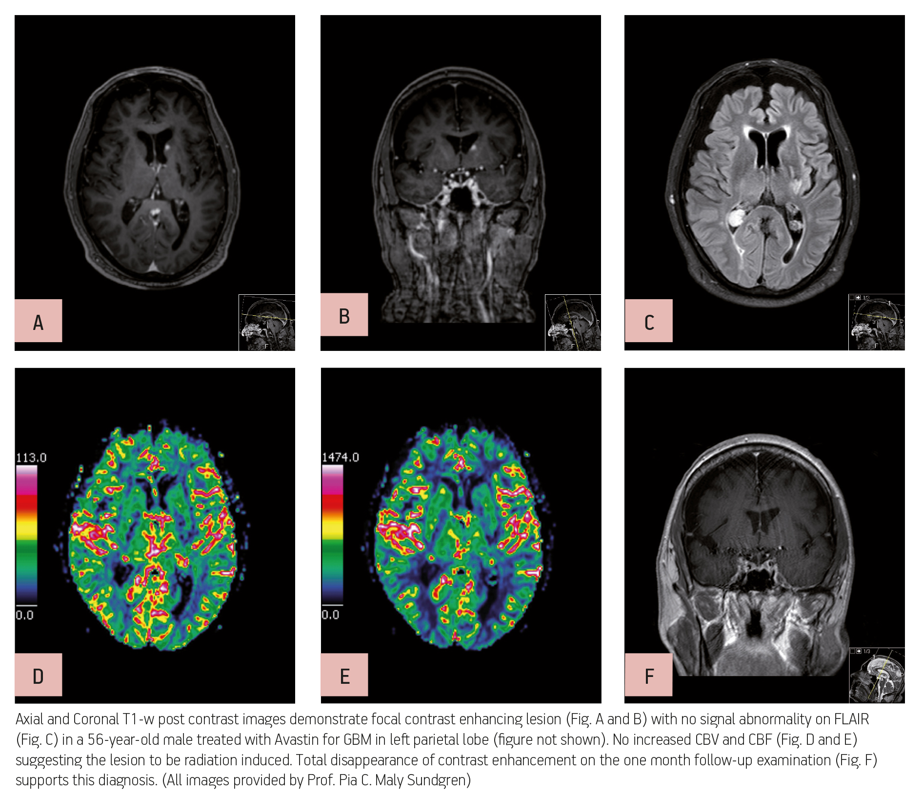
One of the most common types of brain tumour is glioblastoma, which is highly malignant and has a high cell reproduction rate due to the fact that it is nourished by a large network of blood vessels. According to the American Brain Tumour Association there are two types of glioblastoma: primary glioblastomas, which tend to form and make their presence known quickly by growing aggressively, and secondary glioblastomas, which are also aggressive but show slower growth and only represent 10% of all diagnoses.
Read more…
Watch sessions from this Categorical Course on ECR Live:
Imaging the arteries is a daily task for interventional radiologists like José Ignacio Bilbao, president of this year’s ECR, but other subspecialists should know about the main clinical problems associated with the blood vessels. The Clinical Lessons for Imaging Core Knowledge (CLICK) courses, starting this Saturday and finishing Monday, will present delegates with various clinical scenarios and state-of-the-art techniques for imaging vascular disease.
Cardiovascular events still account for the majority of deaths worldwide. Diabetes and hypertension are well-known risk factors that should be monitored and treated appropriately. But the combination of metabolic and cardiovascular factors should also be included in the equation, according to Lars Lönn, professor of vascular surgery and radiology at the National Hospital in Copenhagen. “Cardio-metabolic factors such as obesity are a major risk nowadays; for instance overweight people have a tendency to get fat in the liver, which also gives you a risk for diabetes,” said Lönn, who will chair the course, ‘How old are you in reality? Vascular age and clinical events,’ today at the ECR. Considering these factors is fundamental since the incidence of obesity will continue to rise in the near future, and with it the number of potential cardiovascular complications.
Read more…
Watch this session on ECR Live: Friday, March 8, 16:00–17:30, Room C
Osteoarthritis, a degenerative joint disease, affects a large number of people worldwide. But with the emergence of new MRI techniques, researchers believe they will be able to prevent its development in the near future. Experts will present the latest methods to assess cartilage tissue quality at a very early stage and discuss remaining challenges, in a dedicated New Horizons Session, today at the ECR.
Cartilage is composed of collagen and glycosaminoglycans (GAG), which are responsible for the biomechanical properties of cartilage tissue. An interesting way to image cartilage is to look at the amount of GAG, which decreases at the onset of tissue degeneration, a process which occurs due to ageing or an induced defect, for instance trauma or surgical intervention in the joints. If left untreated, a tissue defect can lead to osteoarthritis. GAGs are known to be among the earliest biomarkers of cartilage degeneration, and if a focal reduction in the amount of GAG can be identified, then therapy to avoid further damage can begin.
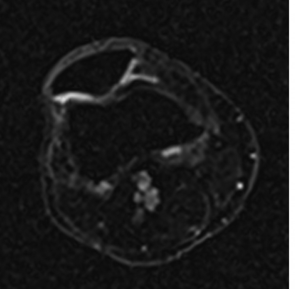
Sodium image in the axial plane of the patella shows the patellar cartilage. At the border from the medial to the lateral facet of the patella an area with decreased sodium signal-to-noise ratio (SNR) is visible which corresponds to a decreased content of glycosaminoglycan (GAG) although the cartilage thickness is preserved. This means an early stage of cartilage degeneration in this area with a focal loss of GAG.
(Provided by Prof. Siegfried Trattnig and the MR Centre of Excellence)
Read more…
Watch this session on ECR Live: Thursday, March 7, 16:00–17:30, Room D2
Over the past decade, technological improvements have led to the widespread use of imaging modalities in the prediction, diagnosis and follow-up of coronary disease. Radiologists now have the ability to obtain information on the structure of cardiac muscle with MRI and evaluate cardiac arteries with CT, while hybrid imaging will soon allow them to do both. Cardiac CT will also provide more functional information in the future, and its use will continue to grow. Experts will present the newest and upcoming possibilities of cardiac imaging today at the ECR.
Advances in cardiac CT have brought its use in clinical routine to unprecedented levels. The main reason is that image acquisition optimisation strategies allow radiologists to assess blood vessels with the same efficiency as coronary angiography, non-invasively and almost instantaneously.

Read more…

Intensive care units are special working environments, presenting radiologists with complex cases and patients with severe conditions. Diagnostic imaging examinations and the work of the radiologist have to be adapted towards these special circumstances, which can be one of the biggest challenges when working in an intensive care unit. Today there is a strong need for accurate, clinically relevant radiological input, which often has to be worked out while facing a lack of adequate image material and patients suffering from life-threatening conditions.
Prof. András Palkó from Szeged, Hungary, will chair the session on imaging in intensive care patients.
The ECR 2013 Special Focus Session on imaging in intensive care patients, chaired by ESR Past-President, Professor András Palkó from Szeged Medical School in Hungary, will give an up-to-date overview on the use of common imaging methods in the ICU environment. Special Focus Sessions are clearly aimed at in-depth analysis and the promotion of scientific debate between the speakers and their audience.
“The intensive care unit is a very special environment requiring special expertise from both the technicians and the radiologists working in a technically challenging situation. The patients are typically in very severe conditions, frequently unconscious, and almost always connected to life-support and monitoring equipment,” Prof. Palkó pointed out some of the difficulties of working in an ICU.
As a result of this, the majority of imaging examinations are performed on patients with limited ability to cooperate and often at the bedside. Reports are then typically written with insufficient clinical information, based on technically limited images, even though the need for accurate imaging material and radiological information is even greater than in standard clinical settings.
Read more…
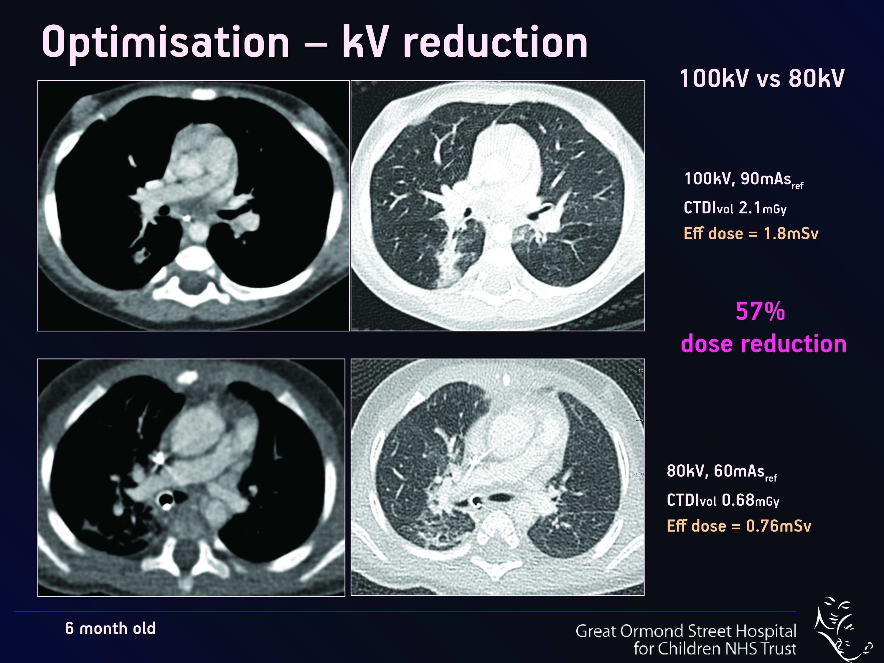
The optimisation and justification of procedures is vital when using CT as an imaging modality; particularly on children, who are more sensitive to ionising radiation than adults. Therefore, it is crucial that all those who use CT understand the physics behind the equipment and ultimately use this understanding to minimise the potential risks while maximising the potential benefits to each individual patient. Patients should also be informed of the risks and benefits of undergoing a CT scan. World-renowned experts will explain these issues in detail during a Special Focus Session at ECR 2013.
“Not all radiologists and technicians are aware of the latest dose reduction strategies. Some are not necessarily so well-informed and perhaps do not realise how important this is. We believe that it is a question of trying to get everybody to a certain level of knowledge and expertise,” said Dr. Catherine Owens, paediatric radiologist and CT unit lead at Great Ormond Street Children’s Hospital in London, U.K.
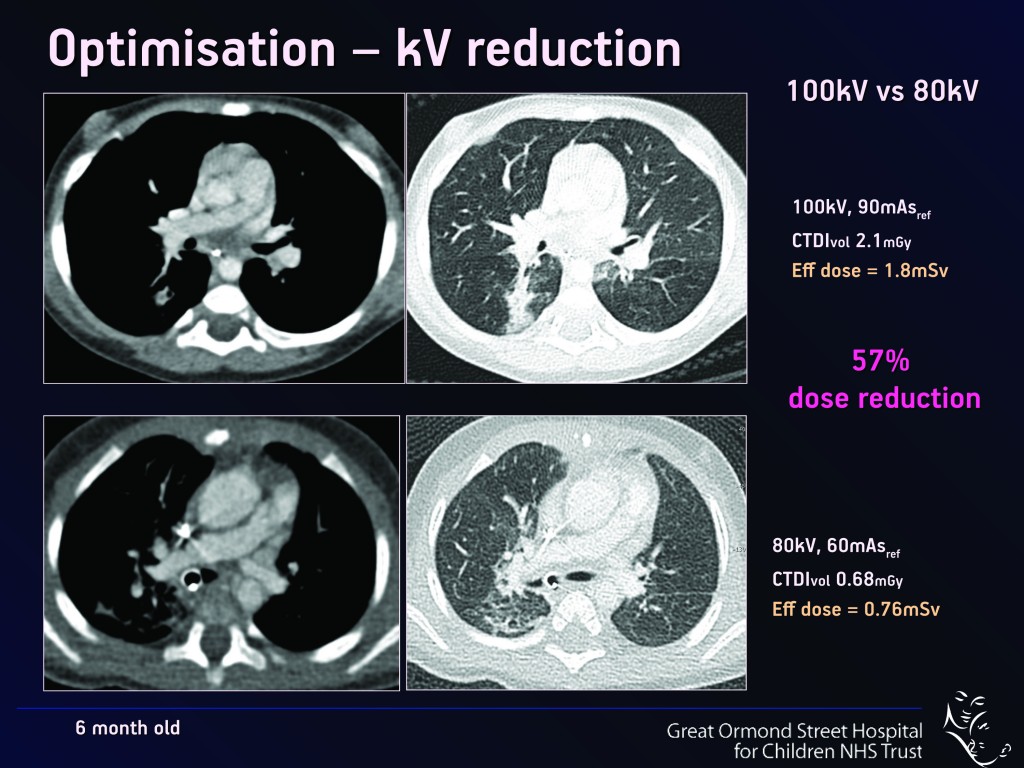
Fig. 1
Read more…
Heart disease affects a very large number of people worldwide, and the consequences can be serious and even lethal. Here, and perhaps more than in many other areas of medicine, imaging has helped to improve treatment and prevention. It does so by detecting the disease at an early stage, sometimes even before its emergence, especially in patients at risk of ischaemic heart disease.
Today, diagnosing cardiac patients has become routine for many radiologists. However, some of them may not know of recent developments in this field and they may need to refresh their knowledge. A panel of experts will update both general and specialised radiologists with the latest information available on cardiac imaging, during the dedicated Mini Course ‘Organs from A to Z: Heart’ at ECR 2013. After an introduction to heart anatomy and the main imaging protocols, the course will focus on valvular diseases and cardiomyopathies; two pathologies commonly encountered in radiology practices.
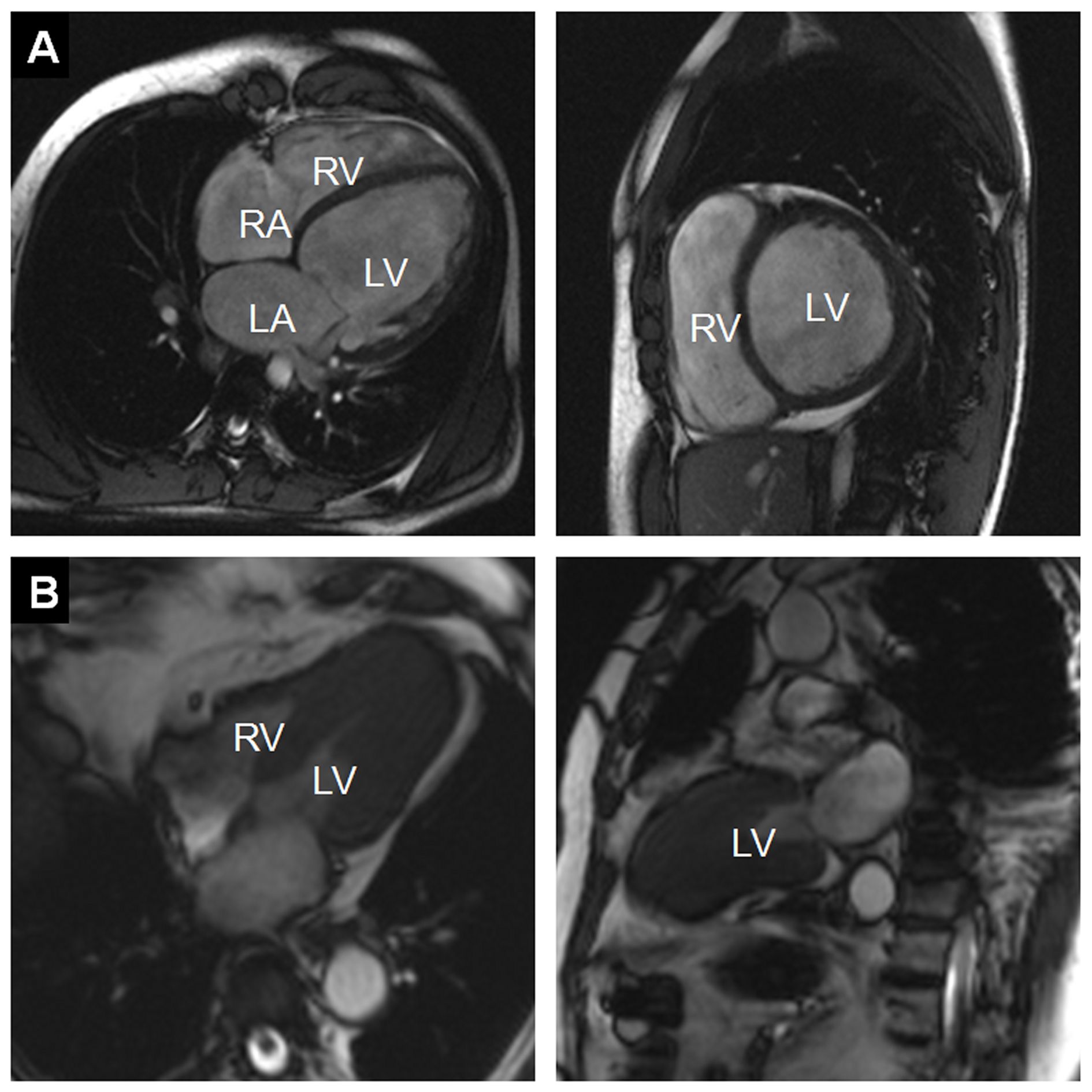
Figure 1:
A) Example of a dilated cardiomyopathy (DCM). Cine-MR images in four-chamber view (left) and short-axis view (right) at end-diastole show significant dilatation of the LV cavity. Ejection fraction was <35% in this patient. (RA = right atrium; LA = left atrium; RV = right ventricle; LV = left ventricle)
B) Example of an asymmetrical, apical hypertrophic cardiomyopathy (HCM). Cine-MR images in a four-chamber (left) and two-chamber view (right) in systole show a markedly thickened left ventricular myocardium predominantly of the apex, as compared with the basal segments (RV = right ventricle; LV = left ventricle).
Read more…