Are we ready to fight a new turf battle in radiation protection?

Recent studies have raised the problem of dose optimisation imaging protocols in patients with renal colic. Some of them are written by emergency physicians, who seem to pay more attention to this problem than radiologists. We would like to hear what you think about this issue in the comments section below.

Renal colic is a common problem, which is increasing in incidence, affects 10%-15% of people over during their lives, and has a tendency to recur. The ability to rapidly identify kidney stones, as well as their position along the ureter and their dimensions, with high sensitivity and specificity using unenhanced CT, has made this technique the first-line approach to the condition. Since CT involves ionising radiation and there is growing concern about its possible carcinogenic effects, low-dose CT protocols for urolithiasis have been developed to minimise radiation risk.

However, low-dose images and often considered as low-quality images and, although these protocols have been shown to be accurate for stone detection, there are concerns about their use due to fears of missing other diagnoses that may clinically mimic stone disease, such as appendicitis, diverticulitis, and cholecystitis. A possible solution could be to use different CT protocols according to the pre-test probability of stone disease. In patients with a previous history of urolithiasis, a low-dose CT examination would be sufficient.
Read more…

Help or put on hold: when do you keep your colleagues waiting?

iStock_000012847617

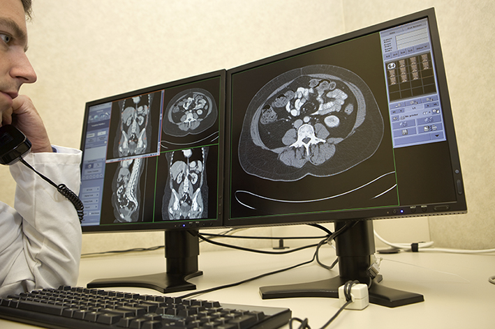
Survey results show that the radiologist’s availability to clinical colleagues is an important part of the imaging service. So how do you balance that availability and your regular workflow? We want to hear your thoughts below.

In 2011, Lindsay et al. published the results of a survey aimed at assessing what factors affected satisfaction with radiology services amongst referring clinicians (1). The survey was conducted shortly after implementation of a PACS system across three hospitals, and focused mainly on the changes created by improved communication between radiology and other hospital departments. Overall, respondents had a positive opinion of the impact of PACS installation, with the majority feeling it decreased the reporting times and ameliorated the working pattern for medical staff. Furthermore, there were a number of indicators showing that direct access to the radiological images did not decrease the importance of the radiology reports. On the contrary, the higher the experience of the referring clinicians, the higher the perceived value of this part of the radiologists’ work.
Read more…

‘Image factories’ or ‘clinical imaging services’: what does the future hold for radiology departments?

A recent article in the New England Journal of Medicine raises some very interesting questions about the future of imaging service provision. Who are we as radiologists, and where do we want to go? Are we running “imaging factories” or “clinical imaging services”? We would like to hear what you think in the comments section at the bottom of this article.

Are modern radiologists just cogs in an image production machine?

Dr. Saurabh Jha is a radiologist working in the Department of Radiology at the Hospital of the University of Pennsylvania, Philadelphia. Before that, he had a professional life as a surgical trainee on this side of the pond, in England. In a paper entitled From imaging gatekeeper to service provider – a transatlantic journey
(N Engl J Med 2013:369:5-7) he underlines the differences between the way he perceived radiologists when he worked in Europe as a surgeon and the way he practices radiology in the USA. English radiologists were gatekeepers: that is, they provided imaging studies only when they were really appropriate and necessary according to their clinical judgement. American radiologists are service providers; that is they perform and read the examinations requested according to the referring physicians’s clinical judgement.

“Evaluation of radiological services in the USA is based on the volume of examinations and turnaround time; the higher the number of studies, the better it is for the department.”

Dr. Jha explains that this difference is mostly related to the fact that imaging was a scarce commodity when he was working for the British National Health Service while, on the contrary, there is abundance of CT scanners, MRI machines, and technologists in the United States. Another explanation is that evaluation of radiological services in the USA is based on the volume of examinations and turnaround time. In such a case, the higher the number of studies, the better it is for the department.
Read more…

Population imaging studies gain ground in healthcare

ECR2015_ECRToday_Blog

Watch this session on ECR Live: Thursday, March 5, 16:00–17:30, Room L1
Tweet #ECR2015L1 #PC8B

Imaging large cohorts of people enables scientists to collect information useful for science and emphasises radiology’s role in healthcare. From the most recently available imaging biomarkers to data such as genomics and metabolomics, Thursday’s dedicated Professional Challenges Session will show just how useful population imaging studies have become in the prognosis of countless diseases.

When radiologists follow a cohort of people for 20 or 30 years, the benefits for the patient increase tremendously. If a radiologist performs a CT examination of a patient’s coronary arteries and finds calcification, chances are that the patient will have a heart attack within the next few years. Unfortunately at this stage, the patient is usually out of the radiologist’s reach.

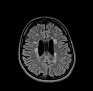
The fluid-attenuated inversion recovery axial image of a 26-year-old female participant shows multiple, bilateral, asymmetric, linear and ovoid hyperintensities that are located perpendicular callososeptal characteristic of multiple sclerosis

The fluid-attenuated inversion recovery axial image of a 26-year-old female participant shows multiple, bilateral, asymmetric, linear and ovoid hyperintensities that are located perpendicular callososeptal characteristic of multiple sclerosis

However, if patients chose to participate in a population study, they will be checked on a regular basis, and radiologists will be able to access previous information and initiate appropriate treatment earlier, significantly
improving patient outcome.

Securing imaging data is always tricky and population imaging studies are an opportunity for radiologists to access this data. Showing the relevance of imaging findings highlights radiology’s role in the medical continuum, according to Prof. Norbert Hosten, of the Ernst-Moritz-Arndt University in Greifswald, Germany, who will chair
the session Thursday.

“Our way to prove that radiology can make people healthier and happier is to do large population imaging studies. Radiology can develop the kind of data that are necessary to prove that our methods really help the patient,” he said.

Read more…

Experts sound the alarm on dangers facing radiologists

ECR2015_ECRToday_Blog

Watch this session on ECR Live: Wednesday, March 4, 16:00–17:30, Room E2
Tweet #ECR2015E2 #PC4A

Remember HAL 9000, the murderous computer in ‘2001: A Space Odyssey’? This scenario doesn’t seem too far off when it comes to radiologists and the constant evolution of their technology. Luckily, they can still stay in control if they change their behaviour and remain at the head of the imaging process, experts will argue during a Professional Challenges session today at the ECR.

Radiologists must claim their place in the medical team if they are to survive. They must come out of the shadows and show that they can bring added value to the team, according to Jim Reekers, professor of interventional radiology at the University of Amsterdam, who will chair the session.

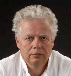
Jim Reekers, from the University of Amsterdam, will chair the session on the role of the radiologist.

Jim Reekers, from the
University of Amsterdam, will
chair the session on the role of the
radiologist.

“I want to make my colleagues aware that they should step out of their offices and act as doctors who work in multidisciplinary teams. Being the photographers or doctors who only give a report with a picture will not secure their future and the reason is very simple: anybody can make and look at a picture nowadays. The difference the radiologist brings is that he or she can give an interpretation of the picture within the context of a patient’s clinical situation. So the radiologist should become a doctor who gets involved with the patient’s situation,” he said.

There is more than one way to do this. For starters, to be on the same level as clinicians, radiologists must behave as such. That means they have to do more than just show the images in multidisciplinary team meetings, they have to sit and discuss them with the doctors.

“Have a junior show the images for you. While my junior presents the pictures, I am sitting together with the other specialists, and suggesting treatment and follow-up options or further diagnostic investigation. It is important to sit with the other doctors. There’s a big difference in how people see me. I’m one of them instead of being the guy who shows the pictures,” Reekers said.

Read more…

Sunday’s sessions for radiographers

ECR2014_ECRToday_Blog_Final

This year’s ECR programme features another great selection of sessions aimed at radiographers. ECR Today spoke to Jonathan McNulty, co-chairman of the ECR 2014 radiographers subcommittee, and Prof. Graciano Paulo, president of the European Federation of Radiographer Societies (EFRS), for their views on the sessions taking place on Sunday.

RC 1214: How important are state-of-the-art displays to radiology?
Watch it on ECR Live: Sunday, March 9, 08:30–10:00, room BRA
Tweet #ECR2014BRA #RC1214

Jonathan McNulty: Most of the technology we use now in Europe is digital, so what this session aims to look at is the state-of-the-art displays we are using today in medical imaging. Their quality is essential so that we continue to be able to pick up on the most subtle anatomical and pathological detail in our images, so the resolution and contrast specifications are important, as are design features that help minimise reflection or glare. A lot of research and design goes into the primary class displays that radiologists use to report from, because that report is vital and can change things dramatically for a patient if a pathology is picked up or missed. There has also been a lot of discussion about handheld devices and the appropriateness of using iPads, other tablets, smartphones or PDAs to view radiology images. Dr. Rachel Toomey, one of the speakers in this session, has done quite a lot of research looking at such devices, which can be very good for reviewing certain types of radiological images but are far from suitable for others.

So this session is going to show what the primary class displays are capable of and why we use them; what the advantages of the more portable devices are and when they can be used appropriately; and then the final presentation will look at quality assurance. Whatever display you use, whether it is a primary class display, a smartphone, or a regular PC monitor, what do we need to keep in mind? What are the quality assurance requirements for clinical use? What do we need to do to on a daily, weekly, or monthly basis to make sure that our displays are not dropping below their peak performance level?

Read more…

ECR 2013 Rec: Role of imaging in personalised therapy monitoring #pc11 #a329

ECR2013Rec_Blog_Cover_A329_PC11

A-329 Role of imaging in personalised therapy monitoring

A.R. Padhani | Saturday, March 9, 16:00 – 17:30 / Room F2

Many imaging biomarkers have emerged which individually or collectively provide unique information on tumour behaviour including response to treatment. There are several requirements that must be met before imaging biomarker(s) can be considered as being able to direct a person’s management. First, the biomarker should have a known biologic basis with a recognised method for quantification and to be adequately validated. With regard to the latter, it is important that the biomarker reports on/measures biologically meaningful cellular/tissue process such as reporting on cell death, on angiogenesis, proliferation and metabolic shutdown NOT simply conveying information on receptor occupancy or down regulation of pathways that may or may not be important. Data acquisition procedures should have been optimised and the test’s performance should have been established. The level of change in the imaging biomarker that can be considered as real should be known (that is, the measurement error). Reproducibility needs to have been determined by appropriately powered test-retest studies. Imaging biomarkers can only be useful if they can detect biologically meaningful effects directly related to treatments (that is, magnitude of biological effects detected must be greater than the reproducibility/measurement error) at appropriate time points to be able to effect patient management. Finally, it must be know how much therapy-induced change is meaningful in terms of patient benefit in terms of hard clinical endpoints such as surgical resectability, organ preservation, progression free and overall survival, etc. These aspects will be considered in detail using practical examples.

ECR 2013 Rec: The Spanish radiographer’s role in advanced MRI research #EM5 #A285

ECR2013Rec_Blog_Cover_EM5_a285

A-285 The Spanish radiographer’s role in advanced MRI research

E. Alfayate Sáez | Saturday, March 9, 14:00 – 15:30 / Room B

A Radiographer, as part of a MRI research team, is more than just a professional obtaining patient´s images for either the investigational studies or clinical trials. Being part of the team means participating and understanding the project as a whole. It means one must know the study’s objectives, collaborate in the protocol design and optimization, and inform the patient about the exam and steps to follow in order to maximize his cooperation. Personal data protection and individual privacy must be guaranteed through all the process; a written informed consent should be signed by the patient as well. Taking care of all these particular aspects is very important for a successful completion of each study/trial. Due to rapid technological advances, and the necessity to deal permanently with state-of-the-art scientific areas, Continuous Professional Development (CPD) for a Radiographer working in a research team is critical. The radiographer is part of a multidisciplinary team, where each professional performs a very specialized task, combining efforts is crucial in order to produce a work of excellence that can be shared with the scientific community. Thanks to the continuous investment in new technology, we have the opportunity, in our site, to conduct research in diverse areas, such as cardiology, traumatology, gynaecology, obstetrics and neurology. Through this presentation we will share some of the research we are working on, as well as the importance of the Radiographer role in a research centre.

ECR 2013 Rec: Finding the time and resources in the radiology department #A024 #PC3

ECR2013Rec_Blog_Cover_PC3_A024

A-024 Finding the time and resources in the radiology department

J. del Cura | Thursday, March 7, 16:00 – 17:30 | Room F1

One of the problems of undergraduate teaching of Radiology is the lack of time for teaching, due to competition for resources with other academic disciplines. Available classroom time and hours of practice are often insufficient to teach the increasingly complex modern Radiology. Also, the availability of financial resources to hire staff or access to educational facilities is competitive and limited, especially in a context of economic crisis. A good solution is to shift the paradigm of education, changing theoretical teaching into self-learning by students. This change allows to free class time to effectively teach Radiology. Classes are converted in workshops, doubt-solving sessions and problem-based learning, all of which matches better with a visual discipline like Radiology. Both on-line classes and e-learning can be useful for this purpose. This kind of teaching also makes Radiology a very attractive discipline for Medicine students. Also, Radiology practices can be carried out using custom computer applications. The lack of professors (and time) for practices can be solved with the help of residents, who are willing to participate as they are more prone to understand the learning needs of the students. Finally, the lack of economic resources makes it is necessary to seek alliances: with the industry, professional associations or with professors from other universities, sharing resources. Internet also provides free materials that can be used to teach.


11
Jul 2013
POSTED BY
DISCUSSION 0 Comments

ECR gives platform to radiographers in ultrasound management debate

ECR2013_ECRToday_Friday_radiographers

Watch this session on ECR Live: Friday, March 8, 16:00–17:30, Room F1

The global demand for medical imaging examinations has been growing rapidly over the past decade. Sustaining a workforce to match demand is becoming a challenge, as an increasing number of hospitals are facing a shortage of radiologists. Some countries have filled the gap by allowing radiographers to perform and interpret ultrasound examinations independently, to relieve the pressure on staff. This option continues to divide the European radiological community, and many seem to be against delegating a medical act to non-doctors. However, new educational opportunities and radiographers’ growing interest in medical science are challenging this concept, a panel of radiographers will show during a Special Focus Session chaired by a radiologist and a radiographer at ECR 2013.

Dean Pekarovic from the University Hospital of Ljubljana, Slovenia.

Dean Pekarovic from the University Hospital of Ljubljana, Slovenia.

Ultrasound is a widely available modality and many medical specialists are using it without the help of radiologists, sometimes without sufficient knowledge and to the detriment of patients. However, radiographers who have received proper theoretical and clinical training know how to best use the modality and read images correctly, according to Dean Pekarovic, a radiographer at the University Hospital of Ljubljana, Slovenia, and member of the advisory board of the European Federation of Radiographer Societies (EFRS). “Ultrasound is a very competitive field, everybody wants to use it. But not everyone has the ability to perform an examination and interpret images appropriately. Radiographers with specific training are able to carry out such examinations and can even write reports on their own,” he said.

Read more…