New treatments give hope to hearing impaired

ECR2015_ECRToday_Blog

Watch this session on ECR Live: Wednesday, March 4, 08:30–10:00, Room E1
Tweet #ECR2015E1 #SF1B

Hearing loss can present many difficulties and obstacles to sufferers, and with ageing populations it’s set to become a major healthcare challenge. Many conditions such as congenital malformation of the inner ear or hypoplastic cochlear nerve can also lead to hearing loss, and sometimes deafness.

Fortunately, many new treatments are available to recover hearing, both partially and completely. Imaging plays an increasingly important role in therapy planning and follow-up, and there is hope on the research front, experts will show during a dedicated Special Focus session on Wednesday morning.
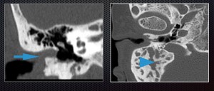
Microtia – congenital anomaly of external and middle ear, resulting in conductive hearing loss. External auditory canal is not patent (arrow), mastoid process is underdeveloped (arrowhead)

Microtia – congenital anomaly of external and middle ear, resulting in
conductive hearing loss. External auditory canal is not patent (arrow),
mastoid process is underdeveloped (arrowhead)

The prevalence of auditory problems in the Western world has doubled over the past 30 years. It is estimated that between 15 and 17% of the population will suffer hearing loss, due to ageing or congenital malformation, but also bad habits, according to Agnieszka Trojanowska, a radiologist at Lublin University Medical School, Poland, who will
chair the session.

“We start to see young adults in their early 30s with sensorineural hearing loss or other related problems because of high frequency noise, which is typical for listening to music. Twenty years ago, such a condition was linked with working in fabrics or on the street. But the good news is that even if you use your iPod a lot, the degree of hearing
loss is light to moderate, so this is not something that will considerably affect your life,” she said.
Read more…

03
Mar 2015
POSTED BY
POSTED IN ,
DISCUSSION 1 Comment

Accomplished neuroradiologist delivers Röntgen lecture at ECR 2014

ECR2014_ECRToday_Blog_Final

In recognition of his exceptional contributions to medical research, particularly in the field of neuroradiology, the European Society of Radiology invited Professor Paul M. Parizel from Antwerp, Belgium, to present the Wilhelm Conrad Röntgen Honorary Lecture at ECR 2014.

Professor Paul M. Parizel from Antwerp, Belgium.

Professor Paul M. Parizel from
Antwerp, Belgium.

Paul M. Parizel is chairman of Antwerp University Hospital’s department of radiology and full professor of radiology at the University of Antwerp’s faculty of medicine. He is also an elected member of the University of Antwerp’s board of trustees, representing the faculty of medicine and health sciences.

In 1982, Prof. Parizel received his medical degree (summa cum laude) from the University of Antwerp and he later went on to earn a PhD degree with a dissertation on ‘The influence of field strength on magnetic resonance imaging: a comparative study in physiochemical phantoms, isolated brain specimens and clinical applications’. He then continued his research thanks to a three-year grant from the Belgian government’s National Foundation for Scientific Research.

Read more…

MRI reveals the human connectome

ECR2014_ECRToday_Blog_Final

Watch this session on ECR Live: Friday, March 7, 16:00–17:30, room BRB
Tweet #ECR2014BRB #NH7

Radiologists often say that the brain is the next frontier. But as diffusion MRI techniques progress, the most mysterious organ in the human body starts to unveil more and more of its secrets, and what was once inconceivable a decade ago is now almost at hand.

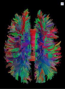
White matter fibre pathways of the brain as depicted with MR tractography. (Provided by Patric Hagmann, CHUV-UNIL, Lausanne, Switzerland)

White matter fibre pathways of the brain as depicted with MR
tractography.
(Provided by Patric Hagmann, CHUV-UNIL, Lausanne, Switzerland)

Researchers are now better able to understand how neurons connect with one another and how disease affects these connections in the human brain. The production and later study of maps of neural connections obtained with MRI are vital to this task. A dedicated New Horizons session will cover this fascinating topic today at the ECR.

Patric Hagmann, who will chair the session, is an attending physician and neuroradiologist at Lausanne University Hospital (CHUV, Centre hospitalier universitaire vaudois) in Switzerland. In his introduction, he will describe what he calls the connectome, a term he coined in his thesis on diffusion MRI and brain connectomics back in 2005*.

“We could sum up the connectome as a comprehensive map of neural connections in the brain. The production and study of connectomes is what we refer to as connectomics; it may range from a detailed map of neurons and synapses within part of, or all of, the nervous system to a description of the functional and structural connectivity between all cortical areas and subcortical structures,” he said.

In his presentation, Hagmann will not only introduce important concepts related to connectomics like scaling, the relation between structural and functional connectivity, and the integration-segregation, but also show how advances in MRI facilitate the mapping of the human connectome.

Read more…

07
Mar 2014
POSTED BY
POSTED IN ,
DISCUSSION 0 Comments

Dr. Pepe’s Diploma Casebook: Case 30 – SOLVED!

Diploma_casebook_case30

Dear Friends,

Showing MRI images of an 82-year-old woman with partial motor seizures afecting her face.

Diagnosis:
1. Metastasis
2. Meningioma
3. Multiple myeloma
4. None of the above

Read more…

Advanced MR imaging helps in interpretation of brain tumours

ECR2013_ECRToday_Cover_Saturday_MRI

Watch this session on ECR Live: Saturday, March 9, 16:00–17:30, Room G/H

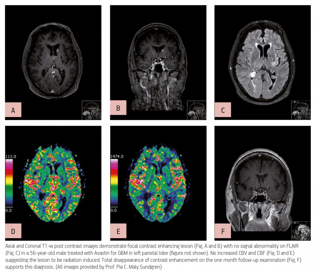
Advanced MR imaging techniques such as perfusion and functional imaging have been a great help in improving the diagnosis and staging of brain tumours. Unlike conventional MR techniques, advanced MR techniques can be used to obtain information not only on the morphological, but also on the functional characteristics of tumours.

One of the most common types of brain tumour is glioblastoma, which is highly malignant and has a high cell reproduction rate due to the fact that it is nourished by a large network of blood vessels. According to the American Brain Tumour Association there are two types of glioblastoma: primary glioblastomas, which tend to form and make their presence known quickly by growing aggressively, and secondary glioblastomas, which are also aggressive but show slower growth and only represent 10% of all diagnoses.

ECRToday_P16_1

Read more…

ECR 2013 Focus: Imaging of the Mind

ECR2013_Focus_7_ImagingMind

The brain remains undoubtedly one of the most mysterious organs of the human body. Magnetic resonance imaging has helped to unveil some of its secrets, and major advances have been made in understanding how the brain functions. Recent developments with resting fMRI (rfMRI) and diffusion MRI (dMRI) indicate that scientists are beginning to see beyond the brain: they have actually started to visualise the human mind. This new information is particularly relevant for understanding complex processes such as dementia, autism and depression. It is also proving increasingly central to the diagnosis of comas and chronic disorders of consciousness.

Leading researchers will discuss where the latest advances have led them and what the future will bring in a dedicated New Horizons Session during ECR 2013. FMRI has been used for over twenty years to visualise changes in brain activity by comparing a task versus a control task, and showing and quantifying how much brain activity is involved in the process. The recent addition of rfMRI enables researchers to track networks that are randomly active. A patient lying in a scanner with no particular task to perform will usually start thinking about the trivialities of the day and go from one thought to the other (“Did I close the door before I left? What am I doing here?” etc.). Neuroresearchers can track this mind mumbling with complex mathematics and extract information from what they call the default mode network.

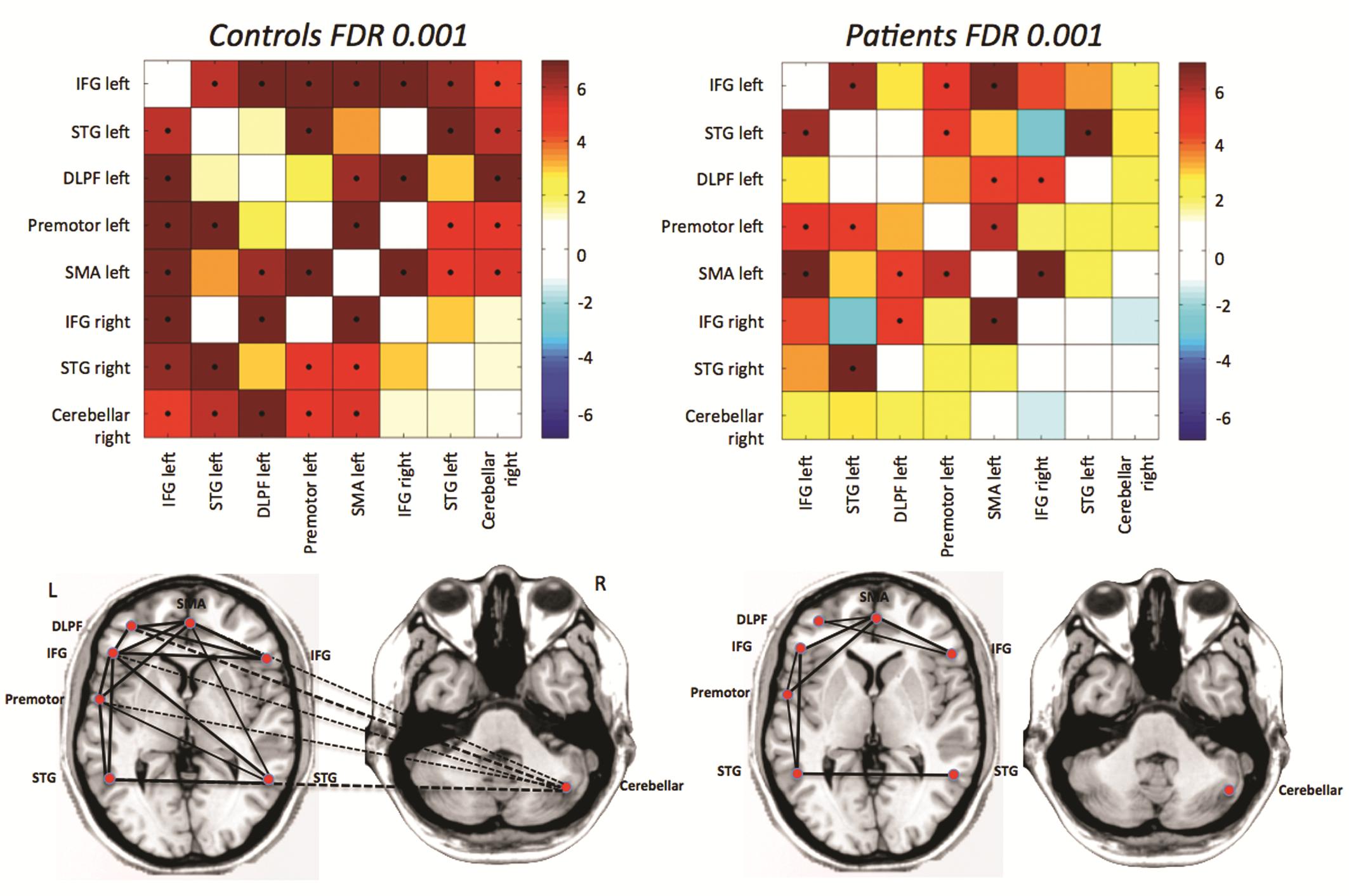
Fig. 1: Differences in functional connectivity from rfMRI between autistic patients and age- and gender matched controls: the major disconnection is between the cerebellum and frontal language areas.

Fig. 1: Differences in functional connectivity from rfMRI between autistic patients and age- and gender matched controls: the major disconnection is between the cerebellum and frontal language areas.

Read more…

Dr. Pepe’s Diploma Casebook: Case 6 – SOLVED!


Dear friends,

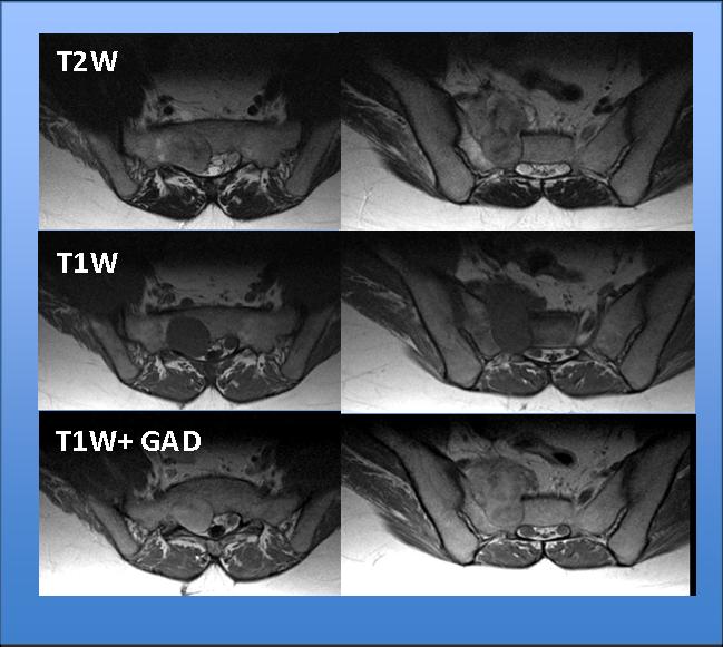
This week I want to show you a neuro/skeletal case, relating to a 25-year-old male with a 3-month history of sciatica.

Fig. 1

Read more…

Dr. Pepe’s Diploma Casebook: Case 2 – SOLVED!

Dear Friends,

Following the usual pattern of an oral examination (mixed cases from all the subspecialities), I challenge you with the following neuro case, below.

Look out for the answer on Thursday. Good luck!

This week’s patient is a 61-year-old immunocompetent male with progressive disorientation and general weakness

Read more…