
Watch this session on ECR Live: Monday, March 10, 08:30–10:00, Room B
Tweet #ECR2014B #SF16a
Shoulder imaging and intervention are becoming more important in clinical practice as ageing populations and patient expectations have increased demand. The shoulder is also one of the joints in the human body that can suffer from a number of pathologic conditions, in both young and elderly patients, such as rotator cuff tears and tendinosis, subacromial-subdeltoid bursitis, calcific tendinopathy, and degenerative conditions.
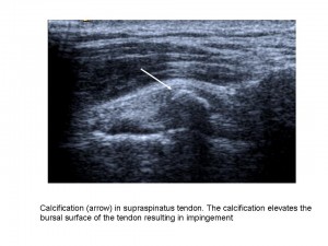
Shoulder imaging and surgery have developed in parallel over the last 20 years, and the introduction of minimally invasive surgical techniques has revolutionised shoulder interventions, which have been facilitated by accurate pre-operative diagnosis. The shoulder is an anatomic area that is very commonly evaluated with musculoskeletal ultrasound as it is accurate, quick, cheap, easily performed, well-tolerated by patients and can be combined with a dynamic examination and interventional procedures.
“MRI provides more general information about the shoulder, but many patients find the examination unpleasant due to noise and pain. Others are excluded from MRI because of claustrophobia or having an embedded electronic device such as a pacemaker. Also MRI cannot be performed as a dynamic examination, it often misses rotator cuff calcification, and the equipment is very expensive. As in many other fields, both techniques rely on high-quality equipment and are operator or interpreter dependent”, said Dr. Ian Beggs, musculoskeletal radiologist at the Royal Infirmary of Edinburgh.
Read more…
Watch this session on ECR Live: Monday, March 11, 08:30–10:00, Room N/O
Tweet #ECR2013NO #SF16B
Organ-sparing surgery and radiation treatment such as intensity-modulated radiotherapy (IMRT) – often combined with chemotherapy – have increased the need for advanced imaging in the head and neck during pre-treament and post-treatment stages. Precision is vital as any tumour that remains undetected outside the treatment field could adversely affect the patients’ prognosis and survival, according to Professor Vincent Vandecaveye, from the department of radiology at the University Hospitals Leuven in Belgium.
It is important to spot any tumour recurrence as early as possible, especially in the post-treatment phase, in order give the patient the best possible chance of salvage treatment. The most common imaging methods in the head and neck area remain CT, MRI and PET-CT; each comes with its own advantages and disadvantages.
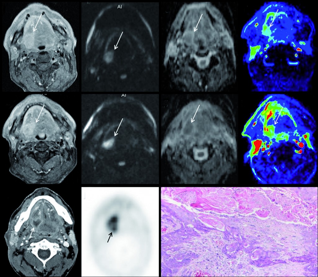
Multiparametric MRI for early treatment prediction of chemoradiation in oropharyngeal cancer: Upper row is pre-treatment MRI of right base of tongue cancer (a=contrast enhanced T1 as anatomical correlate; b=native b1000 diffusion-weighted image; c= ADC-map; d=perfusion-map of IUAC). Middle row is 2 weeks during chemoradiation: same imaging sets, tumour volume will not help. No significant change in b1000, ADC nor perfusion-MRI indicate non-response and thus high risk of tumour relapse after end of treatment. Tumour relapse at PET-CT 8 months after end of treatment, proven by histology (k). (Provided by Professor Vincent Vandecaveye)
Read more…
Watch this session on ECR Live: Sunday, March 10, 08:30–10:00, Room E1
Overuse injuries due to excessive exercise are normally seen in professional athletes, but they are also becoming more frequent in amateur athletes. The Refresher Course on overuse injuries in sports will present three examples of how these injuries, caused by different sports, can be diagnosed and treated.
Gymnastic exercises, for example, are very demanding, not only on the axial but also the peripheral skeleton, and they involve strong forces due to hyperextensive and hyperflexion exercises. A certain degree of hypermobility and increased flexibility is necessary to perform some gymnastic exercises, and so training is required to improve this flexibility. Unnatural movements are sometimes necessary in order to increase this hypermobility and flexibility.
Read more…
Watch this session on ECR Live: Saturday, March 9, 16:00–17:30, Room G/H
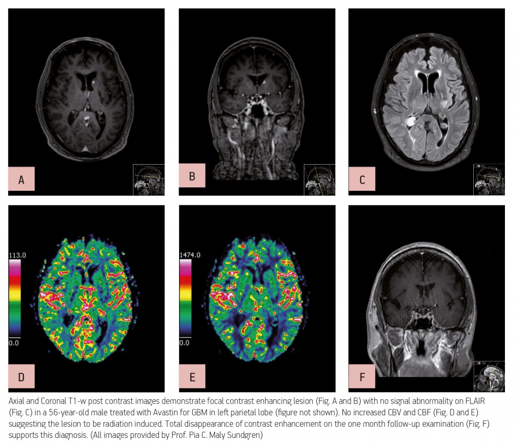
Advanced MR imaging techniques such as perfusion and functional imaging have been a great help in improving the diagnosis and staging of brain tumours. Unlike conventional MR techniques, advanced MR techniques can be used to obtain information not only on the morphological, but also on the functional characteristics of tumours.
One of the most common types of brain tumour is glioblastoma, which is highly malignant and has a high cell reproduction rate due to the fact that it is nourished by a large network of blood vessels. According to the American Brain Tumour Association there are two types of glioblastoma: primary glioblastomas, which tend to form and make their presence known quickly by growing aggressively, and secondary glioblastomas, which are also aggressive but show slower growth and only represent 10% of all diagnoses.
Read more…
Watch this session on ECR Live: Friday, March 8, 08:30–10:00, Room F1
There are a wide range of treatment options available when dealing with hepatocellular carcinoma (HCC), ranging from interventional and endovascular procedures to surgical interventions such as liver transplantation. The main reason for performing endovascular procedures when treating patients with hepatocellular carcinoma is the fact that liver neovascular networks are nourished exclusively by the arteries.
Liver tumours, both primary and metastatic, are almost entirely supplied by branches known as neo-vessels, which originate in the hepatic arteries. The surrounding peritumoural liver parenchyma is vascularised mainly by portal vein branches. When an HCC is larger than two centimetres in diameter the afferent vessel can be identified and then targeted via an arterial endovascular approach. These unique characteristics – dual vascular supply and the ability to identify the afferent vessels – are the rationale behind the use of endovascular treatments, and several different techniques have been developed over the last 30 years. Among the most frequently used are the infusion of chemotherapy and the introduction of particles, either as occluding devices or as carriers of an active agent, which attacks the tumoural cells and surrounding neovessels.

Read more…
Intensive care units are special working environments, presenting radiologists with complex cases and patients with severe conditions. Diagnostic imaging examinations and the work of the radiologist have to be adapted towards these special circumstances, which can be one of the biggest challenges when working in an intensive care unit. Today there is a strong need for accurate, clinically relevant radiological input, which often has to be worked out while facing a lack of adequate image material and patients suffering from life-threatening conditions.
Prof. András Palkó from Szeged, Hungary, will chair the session on imaging in intensive care patients.
The ECR 2013 Special Focus Session on imaging in intensive care patients, chaired by ESR Past-President, Professor András Palkó from Szeged Medical School in Hungary, will give an up-to-date overview on the use of common imaging methods in the ICU environment. Special Focus Sessions are clearly aimed at in-depth analysis and the promotion of scientific debate between the speakers and their audience.
“The intensive care unit is a very special environment requiring special expertise from both the technicians and the radiologists working in a technically challenging situation. The patients are typically in very severe conditions, frequently unconscious, and almost always connected to life-support and monitoring equipment,” Prof. Palkó pointed out some of the difficulties of working in an ICU.
As a result of this, the majority of imaging examinations are performed on patients with limited ability to cooperate and often at the bedside. Reports are then typically written with insufficient clinical information, based on technically limited images, even though the need for accurate imaging material and radiological information is even greater than in standard clinical settings.
Read more…
We see a fair number of odd x-ray images in the ESR office, especially in the search for interesting stories for our Facebook pages. Some can make your stomach turn, some make you laugh, and others just make you think ‘ouch!’ This picture of a foot unnaturally contorted in a high heel shoe naturally caused the latter response…
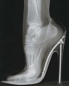
A little digging around on the internet for ‘shoes’ and ‘x-rays’ found a possible source of this image but it also revealed a slightly alarming shady past that radiology and shoes have in common. We’re all aware of the benefits of radiology that nearly everyone gets to experience at least once in their life, but throughout history x-ray technology and radiation have also been used for some, let’s say, ‘questionable purposes’. One of those is certainly the shoe fitting fluoroscope.
Read more…

The WFUMB technical exhibition
This time we didn’t have to travel very far for another episode of our congress coverage, just a couple of stops on the underground and we found ourselves once more at the Austria Center Vienna (ACV), every ESR employee’s second home. But this time there was a difference, 35 degrees outside and the danger of getting sunburn while walking from the underground station to the ACV. So, this is clearly not a story about the ECR, which always takes place in rather more temperate March (and also will take place in March 2012). No, this time nearly the whole ESR staff came to the Austria Center to participate in and help to set up a unique event that has never happened before and will never take place in this form again; the world congress of ultrasound (WFUMB) in conjunction with the EUROSON (congress of the EFSUMB) and the ‘Dreiländertreffen’ (three-country meeting) of Austria, Germany and Switzerland’s ultrasound societies (ÖGUM, DEGUM, SGUM), all held together on 26–29 August, 2011.
One of the big advantages of having our main congress in March is that the ACV is much more comfortable in the cooler times of the year because for some reason it’s easier to heat the building up than to cool it down. On the second day of the congress a big thunderstorm hit Vienna which, besides a lot of rain, brought a welcome drop in temperature. But enough about the weather, after all, our participants came for the congress and its scientific highlights.
Read more…
 Besides hosting the annual ECR in March, the ESR office makes an effort to be present at most of the larger radiology meetings around the globe, whether it’s just around the corner in Hamburg/Germany at the German Radiology Congress or much further away at the Chinese Radiology Congress in Zhengzhou/China. In order to give you an insight into our activities around the world, we hereby present ‘ESR Abroad’. In this first instalment we give you a run-down of our recent visit to the Iranian Congress of Radiology in Teheran/Iran, the annual meeting of the Iranian Society of Radiology (ISR), who participated in the ESR meets programme at ECR 2011.
Before you go to Iran you need to apply for a visa, which can be done either at the local Iranian embassy or directly at the airport in Teheran, but to avoid any difficulties it makes a lot of sense to get a visa before you get on your plane to Iran. We were in the lucky position that two business visa reference numbers were applied for us by the Iranian Society of Radiology so we had no problems applying for visas and entering the country.
Read more…


