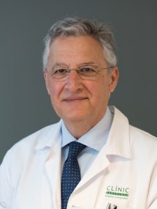
Incoming ESR President lays out clear vision for ECR 2020

By Julia Patuzzi

It is a well-established tradition that on the final day of the congress, ECR Today looks ahead to next year’s ECR. We therefore spoke with Professor Boris Brkljačić from Zagreb, Croatia, the incoming ESR President, who is in charge of ECR 2020. He shared with us some of his ideas and plans for the next European Congress of Radiology.

ECR Today: Professor Brkljačić, the first visual impression of the next congress is always the congress poster. For ECR 2020 you chose artwork by the award-winning Canadian illustrator Peter Diamond, depicting a young woman looking at a small object floating just above her cupped hand. Can you tell us a little about how this particular design came about?
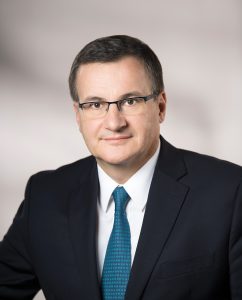
Incoming ESR President Boris Brkljačić is professor of radiology and vice-dean at University of Zagreb School of Medicine, Zagreb, Croatia, and chair of the Department of Diagnostic and Interventional Radiology of University Hospital ‘Dubrava’ in Zagreb.

Boris Brkljačić: The ESR Office provides several options for the congress poster, created by professional designers, and the Congress President and PPC members select one. The selected solution was the best among the proposed options. It resembles Rembrandt’s artwork, with sharp light and dark contrast, and is in good accordance with the slogan for ECR 2020: ‘A Clear Vision for Radiology’. The small floating object represents artificial intelligence, which will be an important topic at the congress, and the names of the ECR 2020 ‘ESR meets’ countries are visible at the bottom of the poster. The 2020 poster contains fewer colours and illustrations compared to the 2019 poster, and is concordant with the visual style of the ESR’s main scientific publication, the journal European Radiology.

ECRT: As the new ESR President, you are also chairperson of the Programme Planning Committee for ECR 2020, which has already been working on preparing the scientific programme for a few months. Can you tell us something about the highlights of the 2020 programme or any specific focus we can expect?

BB: I am very fortunate to have selected excellent Programme Planning Committee members, who are hard-working and dedicated experts in their fields. Planning has already been running at full speed for a few months in order to create a well-balanced programme of very high-quality professional, educational and scientific content. New Horizons Sessions, State of the Art Symposia and Special Focus Sessions have already been selected and mostly created; they are very relevant and balanced, so that young radiologists and experts in particular radiological fields will have interesting sessions to choose from in all areas of radiology. Emerging and hot topics will be covered, like lung cancer screening, artificial intelligence, stroke diagnosis and treatment, and many others. I expect that the plenary/honorary sessions should be the highlight of the congress, as they are interesting for all participants, regardless of their age and expertise. Read more…
How to deal with common diseases in Pakistan: the radiologist’s perspective

by Katharina Miedzinska

Each country has its own special healthcare challenges to shoulder. In Pakistan, a densely populated country located in South Asia, with an estimated population of more than 200 million, major healthcare challenges include exceptionally high prevalence rates for certain diseases. During today’s ‘ESR meets Pakistan’ session, some of the country’s top radiologists will discuss the role radiology plays in managing three of the most common ones: oral cancer, chronic hepatitis and tuberculosis.

Many diseases are common in Pakistan, among them endemic and epidemic infectious diseases, emerging infections, and an increasing burden of non-communicable diseases. The actual burden of infections with the hepatitis B and C virus (HBV, HCV) is approximately seven percent, making Pakistan a country with one of the highest prevalence rates for viral hepatitis in the world. Approximately ten million people in Pakistan are infected with HCV alone. Following the acquisition of the virus, acute HCV infection can progress to chronic infection, which in turn is associated with several morbidities, such as liver cirrhosis and cancer. Besides hepatitis, also malaria, the polio virus (which still circulates in core-reservoirs across Pakistan, although with lesser intensity), dengue outbreaks, and other infectious diseases pose a serious threat to public health security. Over the past few decades, Pakistan has suffered a great deal from these and other infectious diseases. Global warming, changing climate conditions, environmental degradation, and other ecological determinants have a direct effect on these diseases and result in the emergence or re-emergence of infectious entities. The causes of such disease outbreaks are complex and often not well understood.

Read more…

03
Mar 2019
POSTED BY
POSTED IN , ,
DISCUSSION 0 Comments

CEUS ready for clinical paediatric use argues UK expert

Professor Paul Sidhu is Professor of Imaging Sciences at King’s College London and consultant radiologist at King’s College Hospital, where he helped pioneer contrast-enhanced ultrasound (CEUS), a technique that can potentially be used to image children just as well as adults, in reduced time, and without the downsides of radiation and toxicity. Professor Sidhu will make the case for CEUS use in paediatric radiology today during the Luigi Oliva Honorary Lecture.

There is no question that ultrasound is the most child-friendly imaging technique. The modality has countless benefits for young patients: no ionising radiation, no sedation or anaesthesia, and ease of use, to name a few. “Ultrasound is the most suitable modality for imaging children, as they don’t have to stay still. Parents can be present during the examination to support their child. The radiologist can stop and start the examination without losing information, plus children have low body fat and are better suited to ultrasound,” Sidhu said.

Professor Paul Sidhu from King’s College London will argue for CEUS use in paediatric radiology in today’s Luigi Oliva Honorary Lecture.

Adding a contrast agent to ultrasound has been shown to improve diagnosis tremendously in applications where vascularity or haemo-dynamics must be assessed in real time. With CEUS, in the case of focal liver lesions, which are often difficult to characterise in adults and children on B-mode ultrasound alone, essential information from the arterial and portal venous phases aids diagnosis in minutes without needing CT or MR imaging. This alone should be a decisive argument for widespread CEUS use in the paediatric population, Sidhu explained.

“The combination of contrast with ultrasound allows for a more detailed assessment of the pattern of vascularity and dynamics in a continuous fashion for three to five minutes in real time, and it can be repeated as many times as necessary. The child does not need to keep still, and all the same focal liver characteristics seen in the adult are seen in the child. The ability to come to a clear diagnosis, with a single imaging examination is a great relief for the parents, often present with the child during the examination, rather than remote as with CT or MR imaging,” he said.

With CEUS, radiologists can obtain a lot of information in the first five minutes following the injection, the time window that the contrast agent lasts in the blood pool. This is more than enough to carry out the examination and make a conclusion. The technique can be used in every possible scenario from trauma to disease imaging, with great results. “Imaging with ultrasound in children is the most useful and informative procedure,” Sidhu concluded. Read more…

African radiologists call for more cooperation with the ESR in radiation protection

African radiologists will share their knowledge and experience of medical imaging practice in their respective countries, today during the ‘ESR meets Africa’ session. Examples of cooperation with the European Society of Radiology will be presented, notably in radiation protection, a field where efforts between Europe and Africa are starting to pay off.

Africa is big, diverse and full of possibilities. Its myriad of countries (54), languages, cultures and economic scenarios offers unmatched potential, but it can also complicate the organisation of radiology.

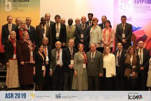
Prof. Dina Husseiny Salama and Prof. Hassen Gharbi, together with ESR President Prof. Lorenzo E. Derchi and Prof. Guy Frija, Chair of EuroSafe Imaging, surrounded by delegates at the 5th African Society of Radiology conference in January 2019, in Cairo, Egypt.

Equipment and workforce tend to vary considerably from one country to another. For example, the radiologists’ ratio ranges from 1 to 80 per million population, depending on the country.

“Our main challenges are in training and the implementation of national legislation that can help organise the field, especially regarding radiation protection,” said Prof. Hassen Gharbi from Tunis, Tunisia, who will co-chair the session with ESR President Lorenzo Derchi from Genoa, Italy.

Technological advances have opened new horizons for the application of ionising radiation in healthcare all around the world and this has led to an increase in medical imaging procedures using radiation, also in Africa.

Unlike most of the continent, Tunisia, Algeria and Morocco have dedicated guidelines on ionising radiation use. But even there, rules must be implemented to help healthcare professionals prescribe examinations adequately.

“There is a growing need for structured strategies and a holistic approach towards the full integration of radiation safety and clinical imaging guidelines in Africa,” said Prof. Dina Husseiny Salama from Cairo, Egypt. Read more…

Systemic effects in interventional oncology: Holy Grail or Pandora’s Box?

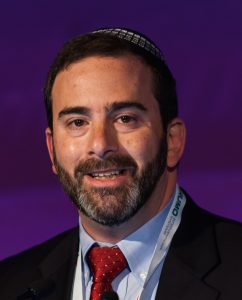
Nahum Goldberg is full professor of radiology at Hadassah Hebrew University, Jerusalem, Israel and visiting professor of radiology at Harvard Medical School, Boston, MA. He is a worldwide expert in image-guided tumour therapy, a field he has helped pioneer and continues to advance. He will share some of the latest results on the effects of these therapies, both positive and negative, today during the Josef Lissner Honorary Lecture.

Image-guided therapy and transcatheter intervention work best on small tumours in the liver, kidney and several other organs, evidence over the past two decades has showed. That was the birth of interventional oncology and it held exciting promise for the (radical) improvement of cancer treatment. Based on that knowledge and promise, many researchers have worked towards combining these procedures with more conventional cancer therapies like chemotherapy and radiation to increase the size and range of the population that can be treated.

Prof. S. Nahum Goldberg from Jerusalem will share some of the latest results on the effects of image-guided tumour therapy, during the Honorary Lecture today at 12:15.

One of the underlined premises of interventional therapies is that using imaging to guide a procedure is less invasive than using other methods like surgery. Despite this and other advantages that have led to ablation systems and techniques now being used clinically more than 100,000 times annually, things have not always gone as planned, according to Goldberg, who heads the interventional oncology unit at Hadassah Medical Centre.

“Specifically, for much of the last two decades, we have argued that other benefits of these interventional oncologic therapies, including percutaneous tumour ablation and chemoembolisation, were focal and local, based upon our conviction that we were only affecting the tumour we were treating and not the entire patient system. It turns out, based on early case reports and more solid evidence produced over the last five years or so, that this premise is not always true,” said Goldberg.

“Most of the damage occurs where intended, but all kinds of pathways in the system are also activated after an intervention. Just like a sunburn that affects a local piece of skin can be accompanied by a headache or fever, a systemic reaction can follow an interventional oncologic procedure in some patients and under certain circumstances. Depending on the type of tumour and its location, a series of both positive and negative systemic effects can be unleashed,” he added.

These effects are rather variable and are currently hard to predict, but one thing is certain: there is a good side and a bad side – the Holy Grail and Pandora’s Box, as Goldberg put it. Read more…

Radiology in Italy takes centre stage on day three of ECR 2019

By Becky McCall

The future status and development of the radiological profession look set to come under the microscope today during the eagerly anticipated ‘ESR meets Italy’ session. The major challenges ahead, including artificial intelligence (AI) and machine learning, are to be addressed by expert speakers.

“We feel that we have to be able to drive AI and not be driven by it. The Italian way of thinking may help our community to fight to ensure that AI does not supersede the human elements of care,” Dr. Corrado Bibbolino, head of the Forensic and Ethical Section of the Italian Society of Medical Radiology (SIRM), told ECR Today ahead of ECR 2019.

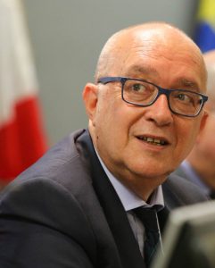
Dr. Corrado Bibbolino is a board member of the Italian branch of Choosing Wisely.

He fears AI may take over the role of the radiologist, and the personal aspects and skills may be lost.

“Patients may think that they are satisfied with AI, but it is not the same as dealing with a real person – it’s not like an automatic ticket machine in a railway station,” Bibbolino said. “Human characteristics like empathy and intuition are not there. A computer might resolve a problem, but it cannot ask the patient questions, feel the reaction, look into the patient’s eyes and think what the patient is thinking – these are things AI cannot do. This is the difference between a human and a robot. AI is important but it is not a substitute for a real radiologist.”

Bibbolino is a long-standing member of the Italian radiology community, having been active in healthcare policy and education for around 40 years. He contributed to the development of national guidelines for radiology practice, including the regulation of teleradiology, and his influence and leadership have led to the establishment of recent Italian laws on healthcare security, professional liability, and insurance reimbursement.

In today’s presentation, ‘Radiology in Italy’, of particular note and relevance will be his work with ‘Choosing Wisely’, which aims to promote dialogue around avoiding unnecessary medical tests and treatments. The initiative is involved with the so-called slow medicine movement, and focuses on a thoughtful, deliberate approach to patient care. Read more…

A new beginning in cancer imaging has just begun, says Beets-Tan

Professor Regina Beets-Tan is chair of the department of radiology at The Netherlands Cancer Institute in Amsterdam, full professor of radiology at the University of Maastricht and adjunct professor of abdominal and oncological radiology at the University of Southern Denmark. She will present the Wilhelm Conrad Röntgen Honorary Lecture, titled ‘Oncologic imaging: a new beginning has just begun’ at ECR 2019.

She shared a few thoughts with us on the future of her specialty in an interview ahead of the congress.

Prof. Regina Beets-Tan from Amsterdam will talk about future aspects of oncologic imaging in today’s honorary lecture.

ECR Today: You have chosen quite an iconic and broad topic for this honorary lecture. What points will you cover exactly?

Regina Beets-Tan: The audience will get a glimpse of the future of cancer care and the role of imaging. The world of cancer medicine is changing rapidly. Major steps forward have been taken. Advanced imaging and computing technology, screening programmes; these all will result in the early detection of more tumours. Minimally invasive treatment, including interventional therapy, will have an increasingly important role. Targeted therapy, which specifically hits the cancer genes, and immunotherapy, which uses the patient’s own immune system to kill cancer cells, will result in prolonged survival of patients who are in the final stage of metastatic disease. It will be ‘precision medicine’; we do not want to give the wrong treatment to the wrong patient. As advocated by Prof René Bernards, a respected leader in cancer research at the Netherlands Cancer Institute: ‘Within 15 years, cancer will become a chronic disease’. And I believe this is true. This transformation will change the way we will practice oncologic imaging. This will require us to recreate our discipline. With this lecture, I would like to take my young colleagues on a 20-minute journey towards their future. Read more…

Welcoming all radiographers to ECR 2019

By Jonathan McNulty, EFRS President

Radiographers have long been involved in the European Congress of Radiology (ECR), and the European Federation of Radiographer Societies (EFRS) has had responsibility for developing the radiographers’ sessions since ECR 2012. However, it is only in recent years that the ECR has become the official scientific congress of the EFRS, and the European Society of Radiology (ESR), for medical imaging radiographers.

Over the past eight congresses, the radiographers’ programme has grown considerably, as has the participation of radiographers. A total of 2,177 radiographers and radiography students, from 75 countries, attended ECR 2018 and we look forward to welcoming even more to ECR 2019, which has now become one of the largest international gatherings of radiographers.

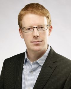
Dr. Jonathan McNulty is Associate Professor and Associate Dean of Graduate Studies at University College Dublin School of Medicine in Dublin, Ireland, and President of the European Federation of Radiographer Societies.

In 2018, a total of 22 refresher courses, professional challenges sessions, special focus sessions, joint sessions, Rising Stars sessions, MyT3 sessions, and scientific sessions made up the radiographers’ programme. For ECR 2019, this will rise to 28 sessions, which will truly offer something for everyone. A special word of thanks must go to Dr. Andrew England from the University of Salford, UK, and a member of the EFRS Educational Wing Management Team, and Dr. Maríanna Garðarsdóttir from Landspitali University Hospital, Iceland, who are the co-chairs of the 2019 radiographers’ scientific subcommittee, and to their team for an excellent educational and scientific programme. Aside from the above sessions, we also look forward to the radiographers’ Voice of EPOS sessions, the involvement of radiographers in a series of sessions at the Cube 2.0 (a special programme dedicated to interventional radiology), and the EFRS Educational Wing annual meeting and our student session.

At ECR 2019, Room C on the 2nd level will become the new venue for most of the sessions in the radiographers’ programme. The Radiographers’ Lounge has also been relocated to Foyer C (outside Room C). In this area, the EFRS and ESR will welcome 20 national radiographers’ societies, along with some educational institutions, who are members of the EFRS Educational Wing, who will all have booths in this area. The radiographers’ Voice of EPOS stage will also be located in the lounge area, as will a number of research studies, requiring your participation, which will take place in the EFRS Radiographers’ Research Hub (Room 2.96). The Radiographers’ Lounge will thus be a great meeting place and, together with the rest of the EFRS Executive Board, I look forward to meeting you in this area and seeing you at the radiographers’ sessions. Read more…

Expand your role and show value, Donoso urges radiologists

Prof. LLuís Donoso-Bach is chairman of the diagnostic imaging department at the Hospital Clínic of Barcelona and professor of radiology at the University of Barcelona. A true leader in European radiology, he has tackled many areas of radiology practice, including liver imaging, digital imaging, IT use in diagnostic radiology, and product development. He has also held the reins of the European Society of Radiology, serving as its president from 2015 to 2016, and sat on various ESR and ECR committees for many years.

Donoso, who is the newly appointed president of the International Society of Radiology, believes the future has much in store for radiology, as he explained ahead of receiving the ESR Gold Medal, the highest honour bestowed by the society, today at the ECR.

Prof. LLuís Donoso-Bach from Barcelona, a former ESR President, will receive the ESR Gold Medal, the highest honour bestowed by the society, today at the ECR.

The role of the radiologist, once marginal in healthcare, has become central in the provision of care, with the introduction of technologies like CT, MR, and interventional radiology. “You won’t find a single health facility running without an imaging department today. A patient hardly ever leaves the hospital without having had at least one imaging examination,” Donoso said.

Radiology has struck a balance between the technology and the clinic. It has become a strong discipline, which must continue to get closer to the patient by getting more involved in treatment and prevention, not just diagnostics, if it is to keep on growing.

Radiologists must go beyond detecting lesions and interpreting images, because machines already perform these tasks better than humans. “This role will be put in question in the years to come. I don’t see, in the near future, any possibility for reading images that haven’t already been pre-processed by a computer assisted detection and interpretation system.”

The radiologist’s role will rather be to answer clinical questions by integrating the imaging information together with clinical information, and putting it all in context.

Radiologists must include more information in their reports, from genomics and fields other than imaging, and not just issue recommendations. To continue to grow, radiologists must become more than radiologists and convert into “experts in imaging information,” Donoso believes. Read more…

Major interventional radiologist receives ESR Gold Medal

Anna-Maria Belli, Professor of Interventional Radiology at St. George’s Hospital in London, UK, will be presented with the Gold Medal of the European Society of Radiology today. Ahead of the ceremony, she shared her views on which directions interventional radiology should take.

Interventional radiology (IR) procedures initially broke into the field of peripheral arterial disease by opening up blocked arteries and establishing angioplasty as a valid, alternative, minimally invasive therapy for those either unfit or unsuitable for standard bypass surgery. It has now become an accepted therapy, replacing open surgery in many situations and responsible for saving limbs from amputation.

IR specialist Prof. Anna-Maria Belli from London will receive the ESR Gold Medal during the ECR 2019 Grand Opening today at 17:45 in Room A.

As experience and skill with arterial catheterisation advanced, so did arterial embolisation, which is used with the opposite intention from angioplasty by selectively occluding arteries. Initially this was an emergency procedure used to treat life-threatening haemorrhage, making it difficult to train in. However, with its expanded indication in the treatment of vascular tumours, it has become a common elective procedure and it has been one of Belli’s special areas of research during the four decades in which she has practised as an interventional radiologist. “Embolisation is now an alternative treatment which may replace standard surgical options, e.g. in the treatment of fibroids and benign prostatic hyperplasia,” she said.


Non-vascular interventional radiology has also grown in leaps and bounds, particularly in the management of cancer with interventional oncology. “The new technologies being introduced into interventional oncology are amongst the most exciting developments and indicate a very strong future for IR,” she believes. Read more…