
A-162 Upper limb nerve entrapment
D. Weishaupt | Friday, March 8, 16:00 – 17:30 / Room E1
The peripheral nerves of the upper limb are affected by a number of entrapment and compression neuropathies. These syndromes involve the brachial plexus as well as the musculocutaneous, axillary, suprascapular, ulnar, radial and median nerves. Clinical examination and electrophysiological studies are traditionally the mainstay of diagnostic work-up. However, ultrasonography and magentic resononance imaging (MRI) may provide key information about the exact anatomic location of the lesion or may help to narrow the differential diagnosis. In certain patients with the diagnosis of a peripheral neuropathy, imaging using either ultrasononography of MRI may help establish the cause of the condition and provide information crucial for conservative management or surgical planning. In addition, imaging is particularly valuable in compex cases with discrepant nerve functions test results.

B-0786 Evaluation of a new method for the assessment of anterior acetabular coverage and hip joint space narrowing
R. Ferré, E. Gibon, A. Feydy, H. Guerini, R. Campana, N. Zee, C. Bourdet, M. Hammadouche, J.-l. Drapé | Monday, March 11, 10:30 – 12:00 / Room E1
Purpose: The Lequesne’s false profile (LFP) view is commonly used to evaluate hip joint space narrowing (JSN) and anterior acetabular coverage using the vertical-center-anterior margin (VCA) angle. A novel low-dose biplanar slot scanner (SS) allows simultaneous acquisitions of weight-bearing oblique views of both hip joints. The aim was to compare LFP views versus biplanar oblique views obtained by SS.
Methods and Materials: LFP views were obtained on 56 hips on a computed radiography system. On the same hips, simultaneous oblique views of both hips were acquired in the SS, with the patient’s pelvis positioned at 45° from each acquisition plane. Two independent observers measured VCA angle and JSN on each acquisition. JSN was evaluated through the joint space/femoral head diameter ratio. Measurements from both techniques were compared using the student t-test and the Pearson’s correlation coefficient. Interobserver agreement of VCA angle and JSN assessments were calculated with the intraclass correlation coefficient (ICC).
Results: VCA angle was 33.5° (SD = 6.2°) with SS and 35° (5.6°) with LFP views (p<0.05). Pearson correlation coefficient between two techniques was 0.78 (p<0.01). JSN was 0,114 (SD = 0.03°) with SS and 0.108 (SD = 0.03°) with LFP views (p<0.05). Pearson correlation coefficient was 0.85 (p<0.01). ICCs for VCA angle were 0.91 with SS and 0.69 with LFP views. ICCs for JSN evaluation were 0.76 for both SS and LFP views.
Conclusion: SS is a reliable, easy, and low-dose evaluation of JSN and VCA angle despite a slight angular positioning compared with LFP.

B-0313 Muscle elastography in patients affected by multiple sclerosis
G. Illomei, G. Spinicci, M. Arru, M. Marrosu | Friday, March 8, 10:30 – 12:00 / Room E1
Purpose: The purpose of the study was to investigate the use of real time elastography in evaluating the muscle stiffness comparing to Ashworth scale. The aim of the study is to create an elastography score for MS patients.
Methods and Materials: We investigated 101 MS patients, 61 women and 40 men. Disability score assessed by Expanded Disability Status Score of Kurtzke scale ranged from 0 and 8.5 with a mean value of 3.5. In all patients a neurological examination was performed and referred to Ashworth scale score by a neurologist. All patients underwent real time elastography on Esaote my Lab Twice with transducer with a high frequency probe 4-13 MHz. The muscles examined were quadriceps of both legs. The study was a double-blinded one, and it was approved by local Ethic Committee.
Results: There was full concordance between the Ashworth scale evaluation and elastography score. However, patients classified as score 0 in the Ashworth scale can be spitted in 0a (total normality of muscle fibers elasticity) and 0b (initial compromising of muscle fibers elasticity). The main result of the study was the creation of an elastography score of muscle stiffness in MS patients that can be compared with Ashworth scale.
Conclusion: Ashworth scale is at present the only method to evaluate muscle stiffness in MS patients. Our study demonstrated for the first time that it is possible to have an imaging method to assess this clinical examination giving new possibilities to follow the evolving of the disease in these patients.
Dear Friends,
This week’s case features a 78-year-old woman after close reduction of a dislocated hip prosthesis.
Diagnosis:
1. Acetabular cup fracture.
2. Dissociation of bipolar hemiarthroplasty
3. Postoperative foreign body.
4. None of the above
Read more…

Dear Friends,
Muppet is running out of chest cases and wishes to show a bone case instead. Presenting an AP radiograph of both hands of a 59-year-old male with pain in the joints.
Diagnosis:
1. Rheumatoid arthritis
2. Psoriatic arthritis
3. Hyperparathyroidism
4. None of the above
Read more…

Watch this session on ECR Live: Sunday, March 10, 08:30–10:00, Room E1
Overuse injuries due to excessive exercise are normally seen in professional athletes, but they are also becoming more frequent in amateur athletes. The Refresher Course on overuse injuries in sports will present three examples of how these injuries, caused by different sports, can be diagnosed and treated.
Gymnastic exercises, for example, are very demanding, not only on the axial but also the peripheral skeleton, and they involve strong forces due to hyperextensive and hyperflexion exercises. A certain degree of hypermobility and increased flexibility is necessary to perform some gymnastic exercises, and so training is required to improve this flexibility. Unnatural movements are sometimes necessary in order to increase this hypermobility and flexibility.
Read more…

Watch this session on ECR Live: Friday, March 8, 16:00–17:30, Room C
Osteoarthritis, a degenerative joint disease, affects a large number of people worldwide. But with the emergence of new MRI techniques, researchers believe they will be able to prevent its development in the near future. Experts will present the latest methods to assess cartilage tissue quality at a very early stage and discuss remaining challenges, in a dedicated New Horizons Session, today at the ECR.
Cartilage is composed of collagen and glycosaminoglycans (GAG), which are responsible for the biomechanical properties of cartilage tissue. An interesting way to image cartilage is to look at the amount of GAG, which decreases at the onset of tissue degeneration, a process which occurs due to ageing or an induced defect, for instance trauma or surgical intervention in the joints. If left untreated, a tissue defect can lead to osteoarthritis. GAGs are known to be among the earliest biomarkers of cartilage degeneration, and if a focal reduction in the amount of GAG can be identified, then therapy to avoid further damage can begin.
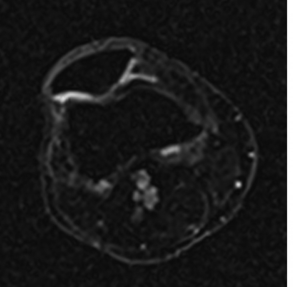
Sodium image in the axial plane of the patella shows the patellar cartilage. At the border from the medial to the lateral facet of the patella an area with decreased sodium signal-to-noise ratio (SNR) is visible which corresponds to a decreased content of glycosaminoglycan (GAG) although the cartilage thickness is preserved. This means an early stage of cartilage degeneration in this area with a focal loss of GAG.
(Provided by Prof. Siegfried Trattnig and the MR Centre of Excellence)
Read more…

Dear Friends,
This week I’m presenting the case of a 9-year-old child with pain in the leg after trauma.
Diagnosis:
1. Aneurysmal bone cyst
2. Simple bone cyst
3. Giant-cell tumour
4. Osteosarcoma
Read more…

Dear friends,
This week I want to show you a neuro/skeletal case, relating to a 25-year-old male with a 3-month history of sciatica.
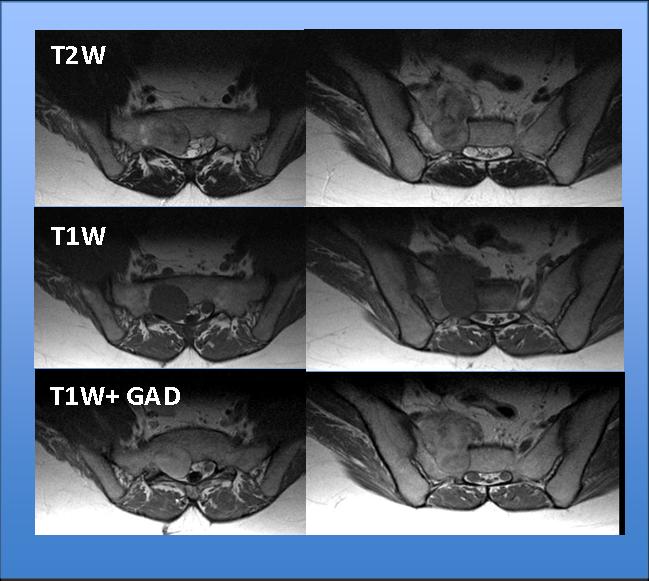
Fig. 1
Read more…
Greece. For some people it’s the perfect destination for their vacation. For some archaeologists it is the perfect place to dig. And for us it was the perfect destination for the Annual Scientific Meeting of the European Society of Musculoskeletal Radiology. Us – that’s Manuela, David, Wolfgang and myself, Maja. The European Society of Musculoskeletal Radiology held its 2011 congress from June 9–11, 2011 in Hersonissos, Crete. It was the first time an ESSR Scientific Meeting has been organised by us – a premiere and therefore quite a big challenge for Manuela and Wolfgang, the main organisers of the event. David and I accompanied them as their personal congress gophers, moral support and Red Bull suppliers.
From the first moment we arrived at the airport in Heraklion we were welcomed by the amazing Greek hospitality, impressively represented by our troubleshooter, Michalis Vlatakis from Summerland Travel, our cooperating travel agent in Crete: a loud “Yassas” (Hello), welcome presents for all, flowers for the ladies and a calming “Nooo problem, this will be the best congress Crete has ever seen” by Michalis. Ευχαριστώ! (Efharisto means “thank you” in Greek)
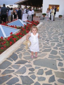
The ESSR takes care of its young academics
Read more…


