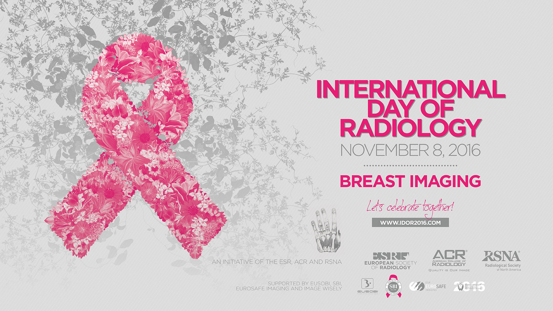
This year, the main theme of the International Day of Radiology is breast imaging. To get some insight into the field, we spoke to Prof. Boris Brkljačić, professor of radiology and Vice-Dean at the University of Zagreb School of Medicine, Croatia, and Chairman of the ESR Communications and External Affairs Committee.
European Society of Radiology: Breast imaging is widely known for its role in the detection of breast cancer. Could you please briefly outline the advantages and disadvantages of the various modalities used in this regard?
Boris Brkljačić: Mammography, ultrasound and MRI are three modalities used for the detection of breast cancer. Mammography has been used for many decades, and the introduction of full flat panel digital mammography has enabled image acquisition with a lower radiation dose, and other advantages in image processing and biopsies. Mammography is used widely in breast cancer screening and has been validated through decades of screening. It is also the initial imaging method in women older than 40 and it enables the detection of microcalcifications, the early signs of ductal cancer in situ, and the majority of breast cancers, depending on the radiographic density of the breast. It can also be used to guide biopsy of microcalcifications. The denser the breasts are, the lower the sensitivity of mammography in detecting breast lesions, which is the disadvantage of mammography. The new mammographic method, digital tomosynthesis, improves the detection rate of cancer in dense breasts. Mammography exposes patients to radiation and is therefore not recommended in young women because their breasts are very radiosensitive.

Prof. Boris Brkljačić, Professor of Radiology and Vice-Dean at the University of Zagreb School of Medicine, Croatia, and Chairman of the ESR Communications and External Affairs Committee.
Ultrasound is an imaging method that provides images based on the acoustic properties of tissues. The blood flow in lesions can be analysed by colour Doppler ultrasound, and elasticity of lesions can be analysed and quantified by sonoelastography. The advantage of ultrasound is that it is completely harmless; it does not expose patients to radiation, and is an excellent method for the guidance of biopsies of all sonographically visible lesions. Ultrasound can demonstrate cancers that are not visible in mammographically dense breasts, and is the complementary imaging modality to mammography, both in diagnosis and in screening. Some U.S. states legally oblige physicians to inform women about mammographic density and advise them of additional methods of examination in dense breasts. Among many advantages in ultrasound technology are the automated whole-breast ultrasound systems that have recently been introduced to the market. The disadvantage of ultrasound is that it increases the number of false-positive findings.
Magnetic resonance imaging (MRI) of the breast has gained considerable importance over the last two decades and is used more and more in breast imaging. It is used in high-risk screening, in the detection of occult cancer with positive lymph nodes, and in the evaluation of implants, and it is the best method for detecting the presence of and assessing the distribution and extent of cancer. It can also be used to monitor the success of neoadjuvant chemotherapy, and is an excellent method for looking for residual cancer or recurrence after treatment. MRI is relatively expensive and time consuming, although abbreviated MRI protocols have recently been introduced.
For treatment planning and monitoring it is very important to know the exact type and grade of cancer, and its immunohistochemical profile. Image guided biopsy is crucial in relation to that, and all imaging methods enable precise, image-guided biopsy to obtain an adequate sample from the breast cancer and other breast lesions.
Read more…