ECR on Demand Preview: Thoracic emergencies #E³ 1520 #A-485

ECROnDemandPreview_Blog_Picture_06

E³ 1520 – Thoracic emergencies, A-458 B – Pulmonary

A short preview of lecture A-458 ‘B. Pulmonary’, from the session E³ 1520 ‘Thoracic emergencies’ at ECR 2014, given by C.M. Schaefer-Prokop from Amersfoort, Netherlands. Watch the whole lecture and many more at http://ipp.myESR.org Direct link: http://bit.ly/Thoracic_emergencies

Sunday, March 9, 16:00 – 17:30 / Room A

Abstract: Acute respiratory failure can have multiple underlying causes including infection, fluid overload, immunological diseases or exacerbation of preexisting lung disease. Since the clinical symptoms are nonspecific, imaging plays an important role. The first imaging method is mostly the chest radiograph, easy to access and to obtain, but non-diagnostic in many cases. (HR)CT offers more possibilities to define the differential diagnosis. The option of this interactive workshop will be to get familiar with the spectrum of diseases that can cause acute respiratory failure and learn about key findings in radiography as well as CT to reduce the differential diagnosis. The interaction between preexisting lung disease, clinical information (e.g. chemotherapy, rheumatoid arthritis, COPD) and imaging findings will be discussed using clinical case studies. Options and also limitations of imaging findings will be illustrated. The following scenarios will be taken into account: acute cardiac failure and various appearances of oedema; acute immunological-toxic disorders including drug-induced lung disease and inhalational injuries; exacerbations of preexisting lung disease including fibrotic and obstructive lung disorders; severe infections causing respiratory failure and their complications.

28
Oct 2014
POSTED BY
POSTED IN ,
DISCUSSION 0 Comments

ECR On Demand Preview: The human connectome #NH 7 #A-158

ECROnDemandPreview_Blog_Picture_05

NH 7 – The human connectome, A-158 – Connectomics in brain pathology (M.P. v.d. Heuvel)

A short preview of lecture A-158 ‘Connectomics in brain pathology’, from the session NH 7 ‘The human connectome: a comprehensive map of brain connections’ at ECR 2014, given by M.P. van den Heuvel from Utrecht, Netherlands.

Watch the whole lecture and many more at http://ipp.myESR.org
Direct link: http://bit.ly/The_human_connectome

Friday, March 7, 16:00 – 17:30 / Room Board Room B

Abstract:

Healthy brain function depends on efficient functional communication within a complex network of structural neural connections, a network known as the connectome. Conversely, damage to the brain’s network, disrupting local neuronal processes and/or global communication between remote functional systems may lead to brain dysfunction. In the last few years, emerging evidence from a wide variety of studies suggests that connectome abnormalities may indeed play an important role in the aetiology of several brain disorders. In my talk, I will discuss the results of recent studies suggesting an important role for affected connectome organization in a number of neurological and psychiatric disorders. In particular, I will highlight the findings of affected functional and structural brain network in neurodegenerative disorders such as Alzheimer’s and ALS, as well as discuss how the application of network science and connectomics may aid our understanding of the biological basis of psychiatric disorders such as autism and schizophrenia.

25
Oct 2014
POSTED BY
POSTED IN ,
DISCUSSION 0 Comments

ECR on Demand Preview: The hand and wrist #RC 1910 #A-585

ECROnDemandPreview_Blog_Picture_04

RC 1910 – The hand and wrist, A-585 C- Tumours and tumour-like lesions

A short preview of lecture A-585 ‘C. Tumours and tumour-like lesions’, from the session RC 1910 ‘The hand and wrist’ at ECR 2014, given by E. Llopis from Valencia, Spain.

Watch the whole lecture and many more at http://ipp.myESR.org

Direct link: http://bit.ly/The_hand_and_wrist

Monday, March 10, 16:00 – 17:30 / Room E1

Abstract:

Radiological study of the wrist and hand is challenging due to its complex anatomy with many small structures and the number of normal bone and soft tissue variants that might mimic injuries. Moreover, many findings can be asymptomatic. Their knowledge is important to avoid misdiagnosis. During this lecture we will also review the role of the different imaging modalities, such as plain films for wrist alignment and bone structures as well as the important role of US and MR in differentiating tumour from tumour-like conditions. We will become familiar with some specific radiological findings that allow us to make accurate diagnoses of soft tissue and bone lesions.

11
Oct 2014
POSTED BY
POSTED IN ,
DISCUSSION 0 Comments

ECR on Demand Preview: RECIST criteria #CC718 #A-187

ECROnDemandPreview_Blog_Picture

CC 718 – Imaging after systemic therapies: the standards, A-187 A. RECIST criteria (Y. Menu)

A short preview of lecture CC 718 ‘Imaging after systemic therapies: the standards’, from the session A-187 ‘A. RECIST criteria’ at ECR 2014, given by Y. Menu from Paris, France.

Watch the whole lecture and many more at http://ipp.myESR.org
Direct link: http://bit.ly/Imaging_after_systemic_…


Friday, March 7, 16:00 – 17:30 / Room Conf. Room M3

Abstract:

The routine practice of oncologic imaging requires standardisation, which means that we need to harmonise technical protocols and agree on the meaning of selected words for the radiological report. The words “Response, “Progression” and “Stable disease” are precisely defined according to internationally accepted thresholds and criteria. Although the rules are quite simple and rather easy to apply, they are very efficient in the classification of the response to treatment, and therefore for the medical decisions. However, the role of the radiologist is not limited to measurements and calculation. The detection of new lesions may be challenging and requires experience. The differential between cancer progression and complications of the treatment might be very difficult and requires an adequate communication with the referring clinician. Overall, most of the decisions taken by the clinician will be related to imaging results, stressing the importance of adequate protocols and reports.

30
Sep 2014
POSTED BY
POSTED IN ,
DISCUSSION 0 Comments

ESR initiatives to strengthen the visibility and role of imaging in personalised medicine

ECR2014_ECRToday_Blog_Final

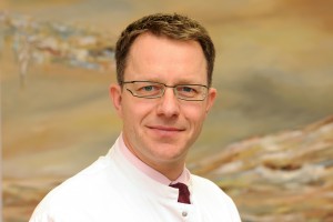
An interview with Prof. Hans-Ulrich Kauczor, Heidelberg/DE, ESR Research Committee Chair

ECR Today: What are the Research Committee’s objectives?
Hans-Ulrich Kauczor: Our main tasks are to periodically survey the needs of the research community, as well as develop recommendations for radiologists to engage with different fields of research and innovation. We also work to assess the current status and develop recommendations on how to improve education in research and to provide strategic recommendations for the research field to the executive council. Last but not least, we aim to leverage cooperation between research disciplines and foster networking and liaising with scientific biomedical European societies.

Hans-Ulrich Kauczor is professor and chairman of radiology at the University of Heidelberg and director of diagnostic and interventional radiology at University Hospital Heidelberg in Germany. He chairs the ESR’s Research Committee.

Hans-Ulrich Kauczor is professor and chairman of radiology at the University of Heidelberg and director of diagnostic and interventional radiology at University Hospital Heidelberg in Germany. He chairs the ESR’s Research Committee.

ECRT: Why is it important to have a dedicated ESR subcommittee for imaging biomarkers?
HUK: The Subcommittee on Imaging Biomarkers was established to address the issues concerning the future development of image-derived quantitative biomarkers, its assessment, validation and standardisation.

The development of imaging biomarkers has become an integral part of modern medicine with a huge potential to advance the development of personalised medicine. Different types of imaging biomarkers (anatomical, functional, and molecular) are used for the detection and treatment of major diseases including cancer, cardiovascular diseases, neurological and psychiatric diseases, musculoskeletal disorders, metabolic diseases, as well as inflammatory and autoimmunity based diseases. In contrast to other biomarkers, imaging biomarkers have the advantage of remaining non-invasive. They are also spatially and temporally resolved, non-destructive and repeatable over a long period, and have the potential for broad application. But before imaging biomarkers can be widely adopted, measures for standardisation and quality assurance must be implemented.

Read more…

Rising Stars exposes students to “the charms of radiology,” says former ECR President

ECR2014_ECRToday_Blog_Final

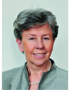
ECR Today spoke with Professor Małgorzata Szczerbo-Trojanowska, from Lublin, Poland, patron of the Rising Stars programme.

ECR Today: The Rising Stars programme is under your patronage this year. What motivated you to take on this role?

Malgorzata Szczerbo-Trojanowska: Medical students and residents will create the shape of radiology in the future. Therefore, those of us who care for our specialty should make an effort to encourage the best, brightest and most enthusiastic students to choose diagnostic imaging as their professional career.

I am very pleased to be involved in the preparation of the Rising Stars programme, which aims to create an interesting educational agenda for students and to encourage them to actively participate in the congress sessions specifically designed for them.

Professor Małgorzata Szczerbo-Trojanowska is Head of the Department of Interventional Radiology and Neuroradiology at the Medical University in Lublin, Poland. She served as ECR Congress President in 2010.

Professor Małgorzata Szczerbo-Trojanowska is Head of the
Department of Interventional Radiology and Neuroradiology at the
Medical University in Lublin, Poland. She served as ECR Congress
President in 2010.

I think this programme is a very important initiative of the European Society of Radiology and the ECR. Having been an academic teacher for many years, I am aware of the great significance of undergraduate education. It has always been my aim as chairperson of the radiology department at my university to offer attractive forms of radiology teaching and to expose students to the charms of radiology from the early years of their education. This is the way to raise their interest in this specialty and get them involved in radiology research. It is in the best interest of the future of radiology. So when I was offered to take patronage of the Rising Stars programme, I didn’t hesitate for even a second.

ECRT: Why should a medical student or radiology trainee take part in this programme?

MST: The European Congress of Radiology, one of the world’s leading radiological meeting, offers medical students and residents a unique opportunity to see the greatest scientists, practitioners, lecturers and teachers present some fascinating achievements of modern radiology. There are also many chances to learn the state of the art in a wide variety of imaging methods for a plethora of diseases, and to find answers to complex problems of contemporary medicine provided by world-renowned experts. Participation in the ECR shows the importance of radiology in patient care, its great impact on other areas of medicine, and its relevance in the progress of medicine.

By taking part in the Rising Stars programme, medical students, radiology trainees and radiography trainees have an opportunity to give a presentation on the results of their own research work and opinions, or ideas, related to medical studies. Visiting the ECR technical exhibition is always an exciting and stimulating experience, providing a unique opportunity to become acquainted with cutting-edge technologies and new solutions.

Read more…

Accomplished neuroradiologist delivers Röntgen lecture at ECR 2014

ECR2014_ECRToday_Blog_Final

In recognition of his exceptional contributions to medical research, particularly in the field of neuroradiology, the European Society of Radiology invited Professor Paul M. Parizel from Antwerp, Belgium, to present the Wilhelm Conrad Röntgen Honorary Lecture at ECR 2014.

Professor Paul M. Parizel from Antwerp, Belgium.

Professor Paul M. Parizel from
Antwerp, Belgium.

Paul M. Parizel is chairman of Antwerp University Hospital’s department of radiology and full professor of radiology at the University of Antwerp’s faculty of medicine. He is also an elected member of the University of Antwerp’s board of trustees, representing the faculty of medicine and health sciences.

In 1982, Prof. Parizel received his medical degree (summa cum laude) from the University of Antwerp and he later went on to earn a PhD degree with a dissertation on ‘The influence of field strength on magnetic resonance imaging: a comparative study in physiochemical phantoms, isolated brain specimens and clinical applications’. He then continued his research thanks to a three-year grant from the Belgian government’s National Foundation for Scientific Research.

Read more…

Award-winning author delivers honorary lecture at ECR 2014

ECR2014_ECRToday_Blog_Final

by Morton A. Meyers

In recognition of his great contributions to abdominal radiology and his insightful publications on the history of science and medicine, Prof. Morton A. Meyers from East Setauket, NY, United States, was invited to deliver the Samuil A. Reinberg Honorary Lecture entitled, ‘The Tempestuous Genesis of MRI: Credit and Discredit’ at ECR 2014.

Prof. Morton A. Meyers from East Setauket, NY, United States.

Prof. Morton A. Meyers from East Setauket, NY, United States.

Prof. Morton A. Meyers is distinguished professor of radiology and medicine, as well as chairman emeritus of the department of radiology at the State University of New York at Stony Brook (SUNY). He is author of the critically acclaimed books; Prize Fight: The Race and Rivalry to be the First in Science and Happy Accidents: Serendipity in Modern Medical Breakthroughs.

His book, Happy Accidents, won the prestigious CHOICE Outstanding Academic Title award in 2009 for excellence in scholarship and presentation, significance of contribution to the field and value as an important treatment of the subject.

In addition to teaching at SUNY, Prof. Meyers has served as visiting professor at more than 70 medical schools around the United States. He has also been in great demand as a lecturer, having received invitations to lecture in Japan, South Africa, China, Israel, Canada, Mexico and throughout Europe, to name but a few places. He has also received a number of awards from national and international societies, as well as the Walter B. Cannon Medal of the Society of Abdominal Radiology. Notably, he delivered the Opening Lecture at ECR 2003, since which he is proud to serve as an Honorary Member of the ESR.

Read more…

13
Mar 2014
POSTED BY
POSTED IN ,
DISCUSSION 0 Comments

Highly-respected Dutch researcher delivers honorary lecture on cardiac imaging

ECR2014_ECRToday_Blog_Final

In recognition of his significant research and work in the areas of MRI, CT and cardiovascular imaging, Professor Albert de Roos from Leiden, the Netherlands, was invited to deliver the Josef Lissner Honorary Lecture entitled, ‘Research in cardiac imaging: how I do it’.

Prof. Albert de Roos from Leiden, the Netherlands.

Prof. Albert de Roos from Leiden, the Netherlands.

Albert de Roos is professor of radiology at Leiden University Medical Center in the Netherlands. Originally from Ermelo, the Netherlands, Prof. de Roos received his medical degree (Cum Laude) from the University of Amsterdam in 1980 and then completed his residency in internal medicine at Zeeweg Hospital Velsen. He then went on to carry out a residency in radiology at St. Elisabeth Gasthuis Haarlem and University Hospital Leiden. In 1985, he was awarded a PhD for his thesis on ‘Biphasic Colon Examination’.

In 1988, Prof. de Roos travelled to the United States to become assistant professor of radiology at the University of Pennsylvania, Philadelphia. He returned to the Netherlands to become associate professor of radiology at Leiden University Medical Center, where he has held a number of posts, including director of magnetic resonance imaging and director of computed tomography. He also served as co-leader of the Interuniversity Cardiology Institute of the Netherlands’ ‘Cardiovascular MRI’ project.

Read more…

EIBIR appoints new scientific director

ECR2014_ECRToday_Blog_Final

by Alena Morrison

Prof. Gabriel P. Krestin has assumed the role of scientific director of the European Institute for Biomedical Imaging Research (EIBIR) after being nominated for the role during the organisation’s General Meeting on Saturday, March 8.

Committed to radiology research, Krestin worked with the ESR to establish EIBIR, which aims to foster and strengthen biomedical imaging research in Europe and has been involved with the organisation since its founding in 2006. He also recently served as chairman of the General Meeting. Since its inception, EIBIR has developed into a key platform for supporting research networking activities, spreading good practice and promoting common initiatives and interoperability in the field of biomedical imaging research. Stakeholders in the EIBIR network have also continued to grow and include European research institutes, shareholder organisations and industry partners.

ECR Today spoke with Krestin to hear some of his thoughts about EIBIR and his ideas for the future of the organisation.

KRESTIN_Gabriel

ECR Today: Looking towards 2014, what activities and initiatives would you like to see begin this year?

Gabriel Krestin: We will have to strengthen the strategy and further extend the activities of EIBIR in the coming years. For now, I hope that at least some of the applications to the first call for proposals in the Horizon 2020 research funding framework of the EC will go into the second phase. That would keep the EIBIR office very busy in the coming months but would also give a new boost to EIBIR’s role as coordinator and administrator of biomedical imaging research in Europe. Furthermore, I hope that some new joint research initiatives will start in image-guided interventions, radiation therapy, paediatric imaging, and neuroimaging. Moreover, the intention is to set up a virtual contract research organisation that will enable multicentre imaging trials, initiated by researchers or by industry, to be performed.

Read more…

13
Mar 2014
POSTED BY
POSTED IN ,
DISCUSSION 0 Comments