Shoulder imaging and intervention

ECR2014_ECRToday_Blog_Final

Watch this session on ECR Live: Monday, March 10, 08:30–10:00, Room B
Tweet #ECR2014B #SF16a

Shoulder imaging and intervention are becoming more important in clinical practice as ageing populations and patient expectations have increased demand. The shoulder is also one of the joints in the human body that can suffer from a number of pathologic conditions, in both young and elderly patients, such as rotator cuff tears and tendinosis, subacromial-subdeltoid bursitis, calcific tendinopathy, and degenerative conditions.

p16_Monday_SF16a_Slide1

Shoulder imaging and surgery have developed in parallel over the last 20 years, and the introduction of minimally invasive surgical techniques has revolutionised shoulder interventions, which have been facilitated by accurate pre-operative diagnosis. The shoulder is an anatomic area that is very commonly evaluated with musculoskeletal ultrasound as it is accurate, quick, cheap, easily performed, well-tolerated by patients and can be combined with a dynamic examination and interventional procedures.

“MRI provides more general information about the shoulder, but many patients find the examination unpleasant due to noise and pain. Others are excluded from MRI because of claustrophobia or having an embedded electronic device such as a pacemaker. Also MRI cannot be performed as a dynamic examination, it often misses rotator cuff calcification, and the equipment is very expensive. As in many other fields, both techniques rely on high-quality equipment and are operator or interpreter dependent”, said Dr. Ian Beggs, musculoskeletal radiologist at the Royal Infirmary of Edinburgh.
Read more…

10
Mar 2014
POSTED BY
POSTED IN ,
DISCUSSION 0 Comments

Experts share strategies to help radiologists justify their imaging decisions

ECR2014_ECRToday_Blog_Final

Watch this session on ECR Live: Sunday, March 9, 16:00–17:30, Room F1
Tweet #ECR2014F1 #SF15B

Evidence-based radiology, comparative effectiveness research and health technology assessment all play a role in the radiologist’s decision-making nowadays. Radiologists must ensure that they are imaging patients at the right time using the right test, and they need to justify radiological examinations and imaging-based interventional procedures on the basis of the best available evidence. The dedicated Special Focus session today at ECR 2014 will teach attendees how to use evidence to justify imaging decisions in day-to-day clinical practice.

The case against overtesting

The case against overtesting

Evidence-based radiology is a relatively new approach designed to inform clinicians of the most appropriate technique to use in a given clinical scenario. It has always been an important topic, but people have recently become more aware of this theme and expect more justification from the physicians, according to Professor Myriam Hunink, who will chair the session. She is a professor of radiology and clinical epidemiology at the Erasmus University Medical Centre in Rotterdam, the Netherlands, and adjunct professor of health decision sciences at Harvard University, Boston, USA.

Read more…

Sunday’s sessions for radiographers

ECR2014_ECRToday_Blog_Final

This year’s ECR programme features another great selection of sessions aimed at radiographers. ECR Today spoke to Jonathan McNulty, co-chairman of the ECR 2014 radiographers subcommittee, and Prof. Graciano Paulo, president of the European Federation of Radiographer Societies (EFRS), for their views on the sessions taking place on Sunday.

RC 1214: How important are state-of-the-art displays to radiology?
Watch it on ECR Live: Sunday, March 9, 08:30–10:00, room BRA
Tweet #ECR2014BRA #RC1214

Jonathan McNulty: Most of the technology we use now in Europe is digital, so what this session aims to look at is the state-of-the-art displays we are using today in medical imaging. Their quality is essential so that we continue to be able to pick up on the most subtle anatomical and pathological detail in our images, so the resolution and contrast specifications are important, as are design features that help minimise reflection or glare. A lot of research and design goes into the primary class displays that radiologists use to report from, because that report is vital and can change things dramatically for a patient if a pathology is picked up or missed. There has also been a lot of discussion about handheld devices and the appropriateness of using iPads, other tablets, smartphones or PDAs to view radiology images. Dr. Rachel Toomey, one of the speakers in this session, has done quite a lot of research looking at such devices, which can be very good for reviewing certain types of radiological images but are far from suitable for others.

So this session is going to show what the primary class displays are capable of and why we use them; what the advantages of the more portable devices are and when they can be used appropriately; and then the final presentation will look at quality assurance. Whatever display you use, whether it is a primary class display, a smartphone, or a regular PC monitor, what do we need to keep in mind? What are the quality assurance requirements for clinical use? What do we need to do to on a daily, weekly, or monthly basis to make sure that our displays are not dropping below their peak performance level?

Read more…

“Cooperation, not competition,” say speakers ahead of ESR Meets ESC session

ECR2014_ECRToday_Blog_Final

Watch this session on ECR Live: Saturday, March 8, 16:00–17:30, Room B
Tweet #ECR2014B #EM3

At each ECR Since 2007, the ‘ESR meets’ programme has included a partner discipline along with the three guest countries, as a way to build formal bridges between the European Society of Radiology (ESR) and other branches of medicine, and to give congress participants an opportunity to learn about something a little different. At ECR 2014, the programme includes a visit from undoubtedly the largest medical discipline to take part in the initiative so far: cardiology, represented by one of the biggest medical societies in Europe, the European Society of Cardiology (ESC). Cardiology has much in common with radiology, but this is the first time that the two European societies have come together for an official joint session at a major meeting.

Read more…

Radiographers sessions on Saturday

ECR2014_ECRToday_Blog_Final

This year’s ECR programme features another great selection of sessions aimed at radiographers. ECR Today spoke to Jonathan McNulty, co-chairman of the ECR 2014 radiographers subcommittee, and Prof. Graciano Paulo, president of the European Federation of Radiographer Societies (EFRS), for their views on the sessions taking place on Saturday.

Read more…

MRI reveals the human connectome

ECR2014_ECRToday_Blog_Final

Watch this session on ECR Live: Friday, March 7, 16:00–17:30, room BRB
Tweet #ECR2014BRB #NH7

Radiologists often say that the brain is the next frontier. But as diffusion MRI techniques progress, the most mysterious organ in the human body starts to unveil more and more of its secrets, and what was once inconceivable a decade ago is now almost at hand.

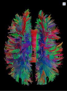
White matter fibre pathways of the brain as depicted with MR tractography. (Provided by Patric Hagmann, CHUV-UNIL, Lausanne, Switzerland)

White matter fibre pathways of the brain as depicted with MR
tractography.
(Provided by Patric Hagmann, CHUV-UNIL, Lausanne, Switzerland)

Researchers are now better able to understand how neurons connect with one another and how disease affects these connections in the human brain. The production and later study of maps of neural connections obtained with MRI are vital to this task. A dedicated New Horizons session will cover this fascinating topic today at the ECR.

Patric Hagmann, who will chair the session, is an attending physician and neuroradiologist at Lausanne University Hospital (CHUV, Centre hospitalier universitaire vaudois) in Switzerland. In his introduction, he will describe what he calls the connectome, a term he coined in his thesis on diffusion MRI and brain connectomics back in 2005*.

“We could sum up the connectome as a comprehensive map of neural connections in the brain. The production and study of connectomes is what we refer to as connectomics; it may range from a detailed map of neurons and synapses within part of, or all of, the nervous system to a description of the functional and structural connectivity between all cortical areas and subcortical structures,” he said.

In his presentation, Hagmann will not only introduce important concepts related to connectomics like scaling, the relation between structural and functional connectivity, and the integration-segregation, but also show how advances in MRI facilitate the mapping of the human connectome.

Read more…

07
Mar 2014
POSTED BY
POSTED IN ,
DISCUSSION 0 Comments

American and European emergency radiologists compare experiences

ECR2014_ECRToday_Blog_Final

Watch this session on ECR Live: Friday, March 7, room F1
Four separate sessions, starting 08:30, 10:30, 14:00, and 16:00
Tweet #ECR2014F1 #MC422 #MC522 #MC622 #MC722

For the fourth year in a row, the European Society of Radiology (ESR) and the Radiological Society of North America (RSNA) will hold a joint course on a hot topic in radiology. After oncologic imaging for the past three years, emergency radiology will be under the scope of North American and European radiologists, who will share and compare their experiences in the dedicated Mini Course today at the ECR*.

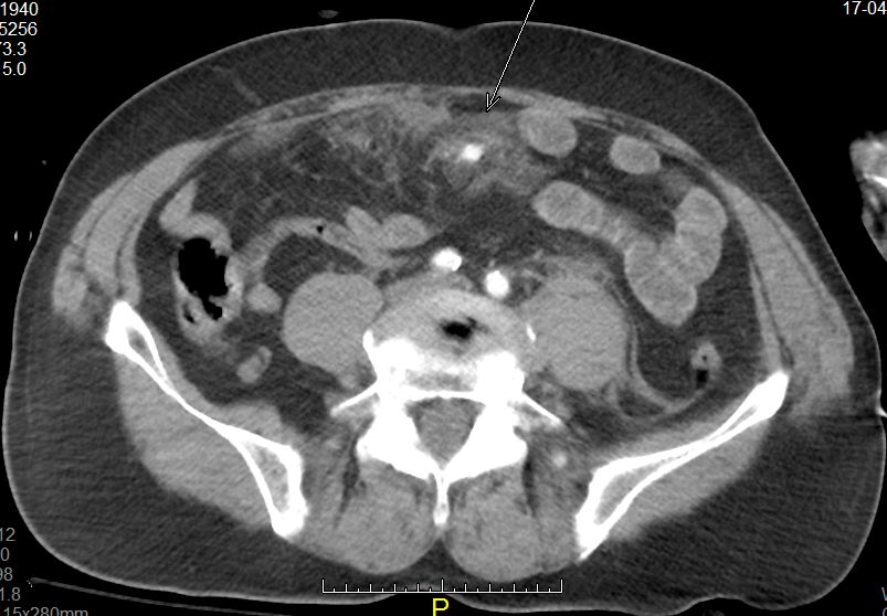
MDCT is the best modality for identifying lesions as subtle as active mesentric bleeding and infiltration (arrow) caused by blunt abdominal injury (lap-belt compression). (Provided by András Palkó)

MDCT is the best modality for identifying lesions as subtle as active mesentric bleeding and infiltration
(arrow) caused by blunt abdominal injury (lap-belt compression). (Provided by András Palkó)

Medical emergencies and trauma are responsible for high levels of morbidity and mortality in all age groups worldwide, and trauma is the leading cause of death in people younger than 45. Consequently, the use of imaging in emergency departments has been increasing exponentially since the 1990s. The demand for CT examinations from emergency departments is, for instance, growing annually by 5% to 15%, depending on the institution.

Even with all the advanced tools imaging has to offer, trauma remains a challenge for most emergency services, according to Ulrich Linsenmaier, associate professor of radiology and head of the department of diagnostic and interventional radiology at KMPP Clinics in Munich, Germany. “Even for advanced level 3 medical centres running their own emergency radiology unit, it is a challenge to integrate advanced radiology services into an interdisciplinary team treating patients with acute traumatic and non-traumatic emergencies,” he said.

Read more…

07
Mar 2014
POSTED BY
POSTED IN ,
DISCUSSION 2 Comments

Radiologists must be thorough when investigating malignant primary bone tumours

ECR2014_ECRToday_Blog_Final

Watch this session on ECR Live: Thursday, 16:00–17:30, Room E2
Tweet #ECR2014E2 #MS3

Malignant primary bone tumours like osteosarcoma and Ewing’s sarcoma are very serious diseases mainly affecting children and teenagers. General radiologists are not likely to see these patients every day at their practice, but when they do, they must know what they have to do to optimise patient care and improve outcomes. Experts will give instructions and share useful advice during the dedicated Multidisciplinary Session today at the ECR.

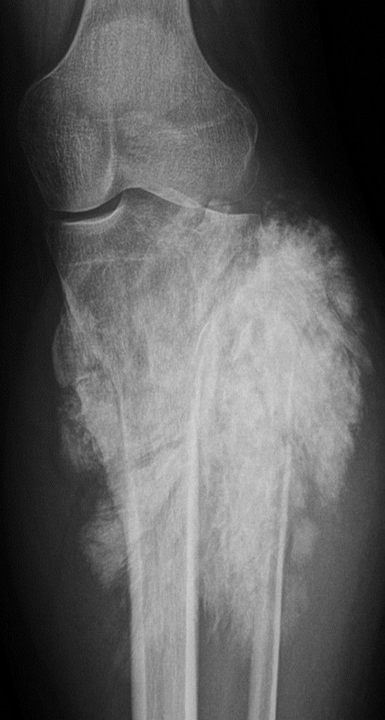
Conventional x-ray of a tumour in the knee (Image provided by Prof. Koenraad Verstraete)

Conventional x-ray of a tumour in the knee
(Image provided by Prof. Koenraad Verstraete)

Read more…

Experts explain how to avoid pitfalls in FDG PET/CT imaging

ECR2014_ECRToday_Blog_Final

Watch this session on ECR Live: Thursday, March 6, 16:00–17:30, Room I/K
Tweet #ECR2014IK #SF3

The demand for PET/CT studies is increasing and so is the need for radiologists to improve their knowledge of this important modality. One of the many areas that require their attention is the occurrence of pitfalls related to the uptake of Fludeoxyglucose (18F), commonly called FDG, the most commonly used tracer in PET/CT imaging. A dedicated Special Focus session at the ECR will offer attendees useful clues on how to avoid these pitfalls and correctly interpret images.

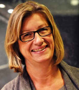
Katrine Åhlström Riklund is director of the medical school and deputy head of the department of radiation sciences at Umeå University, Sweden. She is 2nd vice-chairperson of the ESR’s Congress Committee.

Katrine Åhlström Riklund is director of the medical school and deputy head of the department of radiation sciences at Umeå University, Sweden. She is 2nd vice-chairperson of the ESR’s Congress Committee.

FDG uptake by tissue is also a marker for glucose uptake, which is closely correlated with certain types of tissue metabolism. This means that FDG can show not only disease-related changes but also normal, healthy metabolic changes in the body. “Not everything that shines is pathological. To know the difference, you have to train and learn what is really a disease and what is the physiological distribution of this tracer,” said Professor Katrine Åhlström Riklund, a radiologist specialised in nuclear medicine at Umeå, Sweden, who will moderate the session.
To help radiologists, speakers will share advice regarding FDG uptake in oncology, neurology and cardiology.

Most FDG PET/CT studies are currently being carried out to help stage cancer, and plan and follow-up therapy. The combination of FDG and PET/CT imaging is particularly useful in several different malignancies. Because a tumour cell divides rapidly and has a high rate of metabolism, FDG uptake usually corresponds to disease. Once physicians know the extent of the disease, they can make a more accurate diagnosis and treatment plan, especially in targeted therapies.

Read more…

06
Mar 2014
POSTED BY
POSTED IN ,
DISCUSSION 0 Comments

Next year’s ECR set to be Russian Affair

ECR2013_ECRToday_monday_ECR2014

ECR Today spoke with the next ECR president, Prof. Valentin Sinitsyn. He is chief of the radiology department at the Federal Centre of Medicine and Rehabilitation in Moscow, Russia, and currently serves as president of the European Society of Cardiac Radiology (ESCR).

ECRT: What will be the main highlights of ECR 2014’s scientific programme?

Valentin Sinitsyn: Prepare for more interactivity. People are increasingly interested in interactive sessions to assess what they have learned from a lecture. The ECR has been developing interactive sessions for several years now, but we want to increase that. Today, you can find a lot of information on the internet and many people might not think it is necessary to travel to a congress. We want to create something attractive and show that it is worth coming here. Nothing can replace shaking hands with your colleagues from other countries. I would be very sad if the ECR were entirely online. This is why we are making live meetings more interactive.

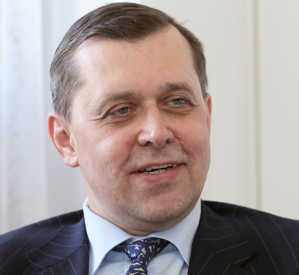
ECR 2014 Congress President Valentin Sinitsyn, from Moscow, Russia.

ECR 2014 Congress President Valentin Sinitsyn, from Moscow, Russia.

We would also like to change the format of scientific sessions. Our lectures have the same format they had one or two hundred years ago: a stage and an auditorium. We are currently discussing the concept of a multimedia classroom, a model which was successfully introduced during the last SIRM congress in June 2012. This multimedia classroom offered 60 work stations from different companies with 25 different cases which were discussed at the end. We are currently discussing the structure with Professor E. Neri from Pisa, who was responsible for the scientific programme of that project.

Soon we are going to use smart phones for voting during audience response sessions. But wireless technology has its limits and sometimes networks crash, so it needs a lot of work. Keypads are an old technology but they are very reliable. I am sure that next year, or the year after that, everybody will be able to vote with their own iPads or tablets.

We will also increase the number of multidisciplinary sessions. This is not something we have to do just during the ECR. This year we had the Imaging Biomarker’s Course the day before the congress, which was organised by the European School of Radiology. This will take place again next year with radiation oncology as the topic.

Read more…