Euro-BioImaging builds pan-European research infrastructure

ECR2014_ECRToday_Blog_Final

Euro-BioImaging will become a pan-European research infrastructure for biomedical imaging technologies. The project’s initial preparatory phase aiming at the development of the technical, legal, governmental and framework of Euro-BioImaging will end in mid-2014.

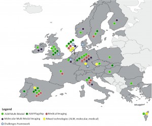
The results of the first Open call for Nodes

The results of the first Open call for Nodes

During the last year of the preparatory phase, Euro-BioImaging successfully carried out the first call for Nodes and finalised plans for its infrastructure model and user access policies.

In spring 2013, Euro-BioImaging published the first open call for Nodes, taking concrete steps towards the construction of a coordinated, open-access imaging infrastructure. Euro-BioImaging invited imaging facilities to submit their expressions of interest in becoming Euro-BioImaging Nodes. In total, 71 proposals for Euro-BioImaging Nodes were submitted by 221 institutions from 19 European countries. Fourteen proposals came from the medical imaging field and presented sound concepts for Nodes on Ultra-highfield MRI, MR/PET, population imaging and phase-contrast imaging. Ten proposals for molecular imaging were submitted.

Read more…

07
May 2014
POSTED BY
POSTED IN
DISCUSSION 0 Comments

ESR initiatives to strengthen the visibility and role of imaging in personalised medicine

ECR2014_ECRToday_Blog_Final

An interview with Prof. Hans-Ulrich Kauczor, Heidelberg/DE, ESR Research Committee Chair

ECR Today: What are the Research Committee’s objectives?
Hans-Ulrich Kauczor: Our main tasks are to periodically survey the needs of the research community, as well as develop recommendations for radiologists to engage with different fields of research and innovation. We also work to assess the current status and develop recommendations on how to improve education in research and to provide strategic recommendations for the research field to the executive council. Last but not least, we aim to leverage cooperation between research disciplines and foster networking and liaising with scientific biomedical European societies.

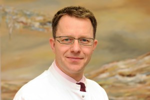
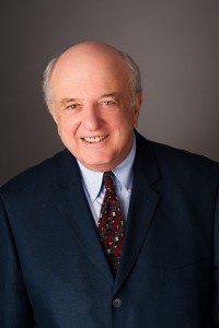
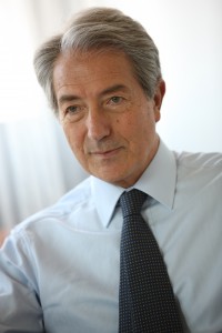
Hans-Ulrich Kauczor is professor and chairman of radiology at the University of Heidelberg and director of diagnostic and interventional radiology at University Hospital Heidelberg in Germany. He chairs the ESR’s Research Committee.

Hans-Ulrich Kauczor is professor and chairman of radiology at the University of Heidelberg and director of diagnostic and interventional radiology at University Hospital Heidelberg in Germany. He chairs the ESR’s Research Committee.

ECRT: Why is it important to have a dedicated ESR subcommittee for imaging biomarkers?
HUK: The Subcommittee on Imaging Biomarkers was established to address the issues concerning the future development of image-derived quantitative biomarkers, its assessment, validation and standardisation.

The development of imaging biomarkers has become an integral part of modern medicine with a huge potential to advance the development of personalised medicine. Different types of imaging biomarkers (anatomical, functional, and molecular) are used for the detection and treatment of major diseases including cancer, cardiovascular diseases, neurological and psychiatric diseases, musculoskeletal disorders, metabolic diseases, as well as inflammatory and autoimmunity based diseases. In contrast to other biomarkers, imaging biomarkers have the advantage of remaining non-invasive. They are also spatially and temporally resolved, non-destructive and repeatable over a long period, and have the potential for broad application. But before imaging biomarkers can be widely adopted, measures for standardisation and quality assurance must be implemented.

Read more…

Rising Stars exposes students to “the charms of radiology,” says former ECR President

ECR2014_ECRToday_Blog_Final

ECR Today spoke with Professor Małgorzata Szczerbo-Trojanowska, from Lublin, Poland, patron of the Rising Stars programme.

ECR Today: The Rising Stars programme is under your patronage this year. What motivated you to take on this role?

Malgorzata Szczerbo-Trojanowska: Medical students and residents will create the shape of radiology in the future. Therefore, those of us who care for our specialty should make an effort to encourage the best, brightest and most enthusiastic students to choose diagnostic imaging as their professional career.

I am very pleased to be involved in the preparation of the Rising Stars programme, which aims to create an interesting educational agenda for students and to encourage them to actively participate in the congress sessions specifically designed for them.
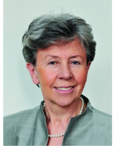
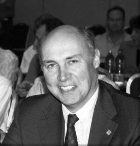
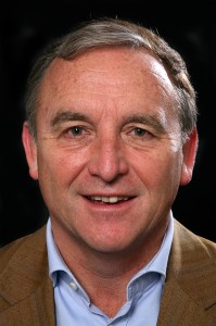
Professor Małgorzata Szczerbo-Trojanowska is Head of the Department of Interventional Radiology and Neuroradiology at the Medical University in Lublin, Poland. She served as ECR Congress President in 2010.

Professor Małgorzata Szczerbo-Trojanowska is Head of the
Department of Interventional Radiology and Neuroradiology at the
Medical University in Lublin, Poland. She served as ECR Congress
President in 2010.

I think this programme is a very important initiative of the European Society of Radiology and the ECR. Having been an academic teacher for many years, I am aware of the great significance of undergraduate education. It has always been my aim as chairperson of the radiology department at my university to offer attractive forms of radiology teaching and to expose students to the charms of radiology from the early years of their education. This is the way to raise their interest in this specialty and get them involved in radiology research. It is in the best interest of the future of radiology. So when I was offered to take patronage of the Rising Stars programme, I didn’t hesitate for even a second.

ECRT: Why should a medical student or radiology trainee take part in this programme?

MST: The European Congress of Radiology, one of the world’s leading radiological meeting, offers medical students and residents a unique opportunity to see the greatest scientists, practitioners, lecturers and teachers present some fascinating achievements of modern radiology. There are also many chances to learn the state of the art in a wide variety of imaging methods for a plethora of diseases, and to find answers to complex problems of contemporary medicine provided by world-renowned experts. Participation in the ECR shows the importance of radiology in patient care, its great impact on other areas of medicine, and its relevance in the progress of medicine.

By taking part in the Rising Stars programme, medical students, radiology trainees and radiography trainees have an opportunity to give a presentation on the results of their own research work and opinions, or ideas, related to medical studies. Visiting the ECR technical exhibition is always an exciting and stimulating experience, providing a unique opportunity to become acquainted with cutting-edge technologies and new solutions.

Read more…

Accomplished neuroradiologist delivers Röntgen lecture at ECR 2014

ECR2014_ECRToday_Blog_Final

In recognition of his exceptional contributions to medical research, particularly in the field of neuroradiology, the European Society of Radiology invited Professor Paul M. Parizel from Antwerp, Belgium, to present the Wilhelm Conrad Röntgen Honorary Lecture at ECR 2014.

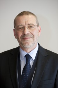
Professor Paul M. Parizel from Antwerp, Belgium.

Professor Paul M. Parizel from
Antwerp, Belgium.

Paul M. Parizel is chairman of Antwerp University Hospital’s department of radiology and full professor of radiology at the University of Antwerp’s faculty of medicine. He is also an elected member of the University of Antwerp’s board of trustees, representing the faculty of medicine and health sciences.

In 1982, Prof. Parizel received his medical degree (summa cum laude) from the University of Antwerp and he later went on to earn a PhD degree with a dissertation on ‘The influence of field strength on magnetic resonance imaging: a comparative study in physiochemical phantoms, isolated brain specimens and clinical applications’. He then continued his research thanks to a three-year grant from the Belgian government’s National Foundation for Scientific Research.

Read more…

Award-winning author delivers honorary lecture at ECR 2014

ECR2014_ECRToday_Blog_Final

by Morton A. Meyers

In recognition of his great contributions to abdominal radiology and his insightful publications on the history of science and medicine, Prof. Morton A. Meyers from East Setauket, NY, United States, was invited to deliver the Samuil A. Reinberg Honorary Lecture entitled, ‘The Tempestuous Genesis of MRI: Credit and Discredit’ at ECR 2014.

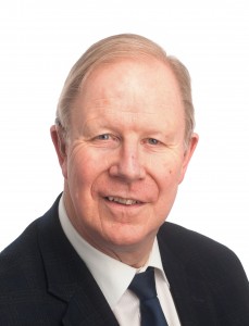
Prof. Morton A. Meyers from East Setauket, NY, United States.

Prof. Morton A. Meyers from East Setauket, NY, United States.

Prof. Morton A. Meyers is distinguished professor of radiology and medicine, as well as chairman emeritus of the department of radiology at the State University of New York at Stony Brook (SUNY). He is author of the critically acclaimed books; Prize Fight: The Race and Rivalry to be the First in Science and Happy Accidents: Serendipity in Modern Medical Breakthroughs.

His book, Happy Accidents, won the prestigious CHOICE Outstanding Academic Title award in 2009 for excellence in scholarship and presentation, significance of contribution to the field and value as an important treatment of the subject.

In addition to teaching at SUNY, Prof. Meyers has served as visiting professor at more than 70 medical schools around the United States. He has also been in great demand as a lecturer, having received invitations to lecture in Japan, South Africa, China, Israel, Canada, Mexico and throughout Europe, to name but a few places. He has also received a number of awards from national and international societies, as well as the Walter B. Cannon Medal of the Society of Abdominal Radiology. Notably, he delivered the Opening Lecture at ECR 2003, since which he is proud to serve as an Honorary Member of the ESR.

Read more…

13
Mar 2014
POSTED BY
POSTED IN ,
DISCUSSION 0 Comments

Highly-respected Dutch researcher delivers honorary lecture on cardiac imaging

ECR2014_ECRToday_Blog_Final

In recognition of his significant research and work in the areas of MRI, CT and cardiovascular imaging, Professor Albert de Roos from Leiden, the Netherlands, was invited to deliver the Josef Lissner Honorary Lecture entitled, ‘Research in cardiac imaging: how I do it’.

Prof. Albert de Roos from Leiden, the Netherlands.

Prof. Albert de Roos from Leiden, the Netherlands.

Albert de Roos is professor of radiology at Leiden University Medical Center in the Netherlands. Originally from Ermelo, the Netherlands, Prof. de Roos received his medical degree (Cum Laude) from the University of Amsterdam in 1980 and then completed his residency in internal medicine at Zeeweg Hospital Velsen. He then went on to carry out a residency in radiology at St. Elisabeth Gasthuis Haarlem and University Hospital Leiden. In 1985, he was awarded a PhD for his thesis on ‘Biphasic Colon Examination’.

In 1988, Prof. de Roos travelled to the United States to become assistant professor of radiology at the University of Pennsylvania, Philadelphia. He returned to the Netherlands to become associate professor of radiology at Leiden University Medical Center, where he has held a number of posts, including director of magnetic resonance imaging and director of computed tomography. He also served as co-leader of the Interuniversity Cardiology Institute of the Netherlands’ ‘Cardiovascular MRI’ project.

Read more…

EIBIR appoints new scientific director

ECR2014_ECRToday_Blog_Final

by Alena Morrison

Prof. Gabriel P. Krestin has assumed the role of scientific director of the European Institute for Biomedical Imaging Research (EIBIR) after being nominated for the role during the organisation’s General Meeting on Saturday, March 8.

Committed to radiology research, Krestin worked with the ESR to establish EIBIR, which aims to foster and strengthen biomedical imaging research in Europe and has been involved with the organisation since its founding in 2006. He also recently served as chairman of the General Meeting. Since its inception, EIBIR has developed into a key platform for supporting research networking activities, spreading good practice and promoting common initiatives and interoperability in the field of biomedical imaging research. Stakeholders in the EIBIR network have also continued to grow and include European research institutes, shareholder organisations and industry partners.

ECR Today spoke with Krestin to hear some of his thoughts about EIBIR and his ideas for the future of the organisation.

KRESTIN_Gabriel

ECR Today: Looking towards 2014, what activities and initiatives would you like to see begin this year?

Gabriel Krestin: We will have to strengthen the strategy and further extend the activities of EIBIR in the coming years. For now, I hope that at least some of the applications to the first call for proposals in the Horizon 2020 research funding framework of the EC will go into the second phase. That would keep the EIBIR office very busy in the coming months but would also give a new boost to EIBIR’s role as coordinator and administrator of biomedical imaging research in Europe. Furthermore, I hope that some new joint research initiatives will start in image-guided interventions, radiation therapy, paediatric imaging, and neuroimaging. Moreover, the intention is to set up a virtual contract research organisation that will enable multicentre imaging trials, initiated by researchers or by industry, to be performed.

Read more…

13
Mar 2014
POSTED BY
POSTED IN ,
DISCUSSION 0 Comments

Former European Radiology editor-in-chief honoured at ECR 2014

ECR2014_ECRToday_Blog_Final

In recognition of his great service to scientific publishing in Europe and his distinguished clinical career, Professor Adrian K. Dixon from Cambridge, UK, was awarded the Gold Medal of the European Society of Radiology at ECR 2014.

Adrian K. Dixon is Master of Peterhouse, the oldest College at Cambridge University, and Emeritus Professor of Radiology, having been head of the department of Radiology for 15 years. He is also an honorary consultant radiologist at Addenbrooke’s Hospital, Cambridge.

Prof. Adrian K. Dixon from Cambridge, United Kingdom.

Prof. Adrian K. Dixon from
Cambridge, United Kingdom.

From an Irish background, he was born in Cambridge where he earned a bachelor’s degree at King’s College. He qualified in medicine after clinical studies at St. Bartholomew’s Hospital London. He then specialised in general medicine, gaining his MRCP in 1974 before deciding to pursue a career in radiology (an appropriate career in view of his profound deafness). He qualified as a radiologist in 1978 and worked in paediatric radiology at Great Ormond Street Hospital, and in computed tomography at St. Bartholomew’s Hospital. In 1979, he became a lecturer at the University of Cambridge’s department of radiology. He earned his doctor of medicine degree for his thesis on computed tomography of the lumbar spine. In 1986, he was elected a Fellow of Peterhouse, where he became director of medical studies.

Read more…

12
Mar 2014
POSTED BY
POSTED IN ,
DISCUSSION 0 Comments

MRI manufacturers demonstrate the latest innovations, upgrades and enhancements

ECR2014_ECRToday_Blog_Final

by John Bonner

MRI has opened up the human body for radiologists to explore with remarkable precision and to gather clinical information of enduring value for physician colleagues, but the liver has so far proved a real diagnostic challenge. A combination of the organ’s complex vasculature and motion effects in free-breathing patients have meant that other modalities have often been chosen instead of MRI, particularly when looking for metastases in oncology cases.

This situation is changing due to the efforts of imaging vendors who have been working on expanding the role of this technology in body imaging. Visitors to the commercial exhibition can inspect a new software application which improves temporal resolution in MR images along with other developments that make scans both safer and more comfortable for patients.

SIEMENS is unveiling a technology that it says will make contrast-enhanced liver imaging fast and robust and allow free-breathing dynamic liver imaging, giving more patients access to high quality MR-based abdominal imaging.

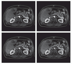
Siemens’ Twist-Vibe MR sequence is designed to enable correct contrast imaging in dynamic liver MRI, allowing fast, robust liver imaging with full 4-D coverage. This series of images shows how the new technology can boost lesion enhancement within the arterial phase: with Twist-Vibe, it is now possible to generate multiple stacks of images from the arterial phase to follow the lesion enhancement over time. (Provided by University Hospital IKRN, Mannheim, Germany)

Siemens’ Twist-Vibe MR sequence is designed to enable correct contrast imaging in dynamic liver MRI, allowing fast, robust liver imaging with full 4-D coverage. This series of images shows how the new technology can boost lesion enhancement within the arterial phase: with Twist-Vibe, it is now possible to generate multiple stacks of images from the arterial phase to follow the lesion enhancement over time. (Provided by University Hospital IKRN, Mannheim, Germany)

Dr. Bernd Ohnesorge, chief executive of Siemens’ MR business unit, explained that the key software technologies underlying the company’s new Twist-Vibe and StarVibe features will be available together as a package called FREEZEit. The former is a sequence that offers high temporal and spatial resolution with full 4D coverage for multi-arterial imaging with 100% precise contrast-timing. Meanwhile, StarVibe is an application that delivers robust, free-breathing, and contrast-enhanced exams for non-compliant patients by resisting motion artefacts.

“These are acceleration techniques that allow us to do body and liver imaging at such high speed that it creates a genuine breakthrough in temporal resolution. So even in an organ as notoriously difficult to image as the liver, we can eliminate motion artefacts while also substantially enhancing contrast timing. Together that makes for a very accurate diagnosis,” he said.

Read more…

12
Mar 2014
POSTED BY
POSTED IN ,
DISCUSSION 0 Comments

New ESR President wants to draw national societies close

ECR2014_ECRToday_Blog_Final

ECR Today caught up with incoming ESR President Professor Lorenzo Bonomo at ECR 2014, and asked him about his vision for the year to come.

ECR Today: What are your plans and ambitions regarding your presidency?
Lorenzo Bonomo: It is with great pride that I take on the presidency of one of the most important and prestigious international scientific societies in the world. I’m also particularly pleased that my presidency coincides with the ESR’s tenth anniversary, which we’ll be celebrating next year.

New ESR President, Prof. Lorenzo Bonomo, from Rome.

New ESR President, Prof. Lorenzo Bonomo, from Rome.

So much has been done over the past few years thanks to the work of my predecessors, the Executive Council, the statutory committees, subcommittees, working groups and the tireless and efficient support of the ESR staff. Many of the objectives in the roadmap approved in 2012 have been achieved together with other new important projects, started during Prof. Frija’s presidency, and it will be my duty to consolidate and bring them to a conclusion. Among them are the development of a clinical decision support system in Europe, the creation of an accreditation council, and a strengthening of relations with other scientific societies in Europe and the EuroSafe Imaging Campaign. The whole learning, education and training field certainly requires close attention and a collaborative vision.

I would also like to draw the national societies closer, by intensifying dialogue with them, trying to understand their needs, and helping them to promote and increase the visibility of radiology. I would like my role to be that of a facilitator supporting the ongoing projects and promoting the start of new activities that committee chairpersons intend to achieve.

Read more…

10
Mar 2014
POSTED BY
POSTED IN ,
DISCUSSION 1 Comment