African radiologists call for more cooperation with the ESR in radiation protection

African radiologists will share their knowledge and experience of medical imaging practice in their respective countries, today during the ‘ESR meets Africa’ session. Examples of cooperation with the European Society of Radiology will be presented, notably in radiation protection, a field where efforts between Europe and Africa are starting to pay off.

Africa is big, diverse and full of possibilities. Its myriad of countries (54), languages, cultures and economic scenarios offers unmatched potential, but it can also complicate the organisation of radiology.
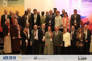
Prof. Dina Husseiny Salama and Prof. Hassen Gharbi, together with ESR President Prof. Lorenzo E. Derchi and Prof. Guy Frija, Chair of EuroSafe Imaging, surrounded by delegates at the 5th African Society of Radiology conference in January 2019, in Cairo, Egypt.

Equipment and workforce tend to vary considerably from one country to another. For example, the radiologists’ ratio ranges from 1 to 80 per million population, depending on the country.

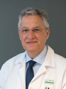
“Our main challenges are in training and the implementation of national legislation that can help organise the field, especially regarding radiation protection,” said Prof. Hassen Gharbi from Tunis, Tunisia, who will co-chair the session with ESR President Lorenzo Derchi from Genoa, Italy.

Technological advances have opened new horizons for the application of ionising radiation in healthcare all around the world and this has led to an increase in medical imaging procedures using radiation, also in Africa.

Unlike most of the continent, Tunisia, Algeria and Morocco have dedicated guidelines on ionising radiation use. But even there, rules must be implemented to help healthcare professionals prescribe examinations adequately.

“There is a growing need for structured strategies and a holistic approach towards the full integration of radiation safety and clinical imaging guidelines in Africa,” said Prof. Dina Husseiny Salama from Cairo, Egypt. Read more…

Systemic effects in interventional oncology: Holy Grail or Pandora’s Box?

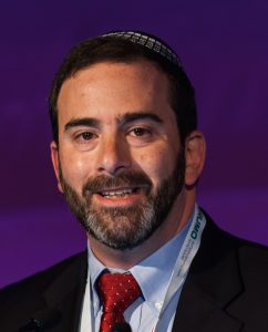
Nahum Goldberg is full professor of radiology at Hadassah Hebrew University, Jerusalem, Israel and visiting professor of radiology at Harvard Medical School, Boston, MA. He is a worldwide expert in image-guided tumour therapy, a field he has helped pioneer and continues to advance. He will share some of the latest results on the effects of these therapies, both positive and negative, today during the Josef Lissner Honorary Lecture.

Image-guided therapy and transcatheter intervention work best on small tumours in the liver, kidney and several other organs, evidence over the past two decades has showed. That was the birth of interventional oncology and it held exciting promise for the (radical) improvement of cancer treatment. Based on that knowledge and promise, many researchers have worked towards combining these procedures with more conventional cancer therapies like chemotherapy and radiation to increase the size and range of the population that can be treated.

Prof. S. Nahum Goldberg from Jerusalem will share some of the latest results on the effects of image-guided tumour therapy, during the Honorary Lecture today at 12:15.

One of the underlined premises of interventional therapies is that using imaging to guide a procedure is less invasive than using other methods like surgery. Despite this and other advantages that have led to ablation systems and techniques now being used clinically more than 100,000 times annually, things have not always gone as planned, according to Goldberg, who heads the interventional oncology unit at Hadassah Medical Centre.

“Specifically, for much of the last two decades, we have argued that other benefits of these interventional oncologic therapies, including percutaneous tumour ablation and chemoembolisation, were focal and local, based upon our conviction that we were only affecting the tumour we were treating and not the entire patient system. It turns out, based on early case reports and more solid evidence produced over the last five years or so, that this premise is not always true,” said Goldberg.

“Most of the damage occurs where intended, but all kinds of pathways in the system are also activated after an intervention. Just like a sunburn that affects a local piece of skin can be accompanied by a headache or fever, a systemic reaction can follow an interventional oncologic procedure in some patients and under certain circumstances. Depending on the type of tumour and its location, a series of both positive and negative systemic effects can be unleashed,” he added.

These effects are rather variable and are currently hard to predict, but one thing is certain: there is a good side and a bad side – the Holy Grail and Pandora’s Box, as Goldberg put it. Read more…

A new beginning in cancer imaging has just begun, says Beets-Tan

Professor Regina Beets-Tan is chair of the department of radiology at The Netherlands Cancer Institute in Amsterdam, full professor of radiology at the University of Maastricht and adjunct professor of abdominal and oncological radiology at the University of Southern Denmark. She will present the Wilhelm Conrad Röntgen Honorary Lecture, titled ‘Oncologic imaging: a new beginning has just begun’ at ECR 2019.

She shared a few thoughts with us on the future of her specialty in an interview ahead of the congress.

Prof. Regina Beets-Tan from Amsterdam will talk about future aspects of oncologic imaging in today’s honorary lecture.

ECR Today: You have chosen quite an iconic and broad topic for this honorary lecture. What points will you cover exactly?

Regina Beets-Tan: The audience will get a glimpse of the future of cancer care and the role of imaging. The world of cancer medicine is changing rapidly. Major steps forward have been taken. Advanced imaging and computing technology, screening programmes; these all will result in the early detection of more tumours. Minimally invasive treatment, including interventional therapy, will have an increasingly important role. Targeted therapy, which specifically hits the cancer genes, and immunotherapy, which uses the patient’s own immune system to kill cancer cells, will result in prolonged survival of patients who are in the final stage of metastatic disease. It will be ‘precision medicine’; we do not want to give the wrong treatment to the wrong patient. As advocated by Prof René Bernards, a respected leader in cancer research at the Netherlands Cancer Institute: ‘Within 15 years, cancer will become a chronic disease’. And I believe this is true. This transformation will change the way we will practice oncologic imaging. This will require us to recreate our discipline. With this lecture, I would like to take my young colleagues on a 20-minute journey towards their future. Read more…

Expand your role and show value, Donoso urges radiologists

Prof. LLuís Donoso-Bach is chairman of the diagnostic imaging department at the Hospital Clínic of Barcelona and professor of radiology at the University of Barcelona. A true leader in European radiology, he has tackled many areas of radiology practice, including liver imaging, digital imaging, IT use in diagnostic radiology, and product development. He has also held the reins of the European Society of Radiology, serving as its president from 2015 to 2016, and sat on various ESR and ECR committees for many years.

Donoso, who is the newly appointed president of the International Society of Radiology, believes the future has much in store for radiology, as he explained ahead of receiving the ESR Gold Medal, the highest honour bestowed by the society, today at the ECR.

Prof. LLuís Donoso-Bach from Barcelona, a former ESR President, will receive the ESR Gold Medal, the highest honour bestowed by the society, today at the ECR.

The role of the radiologist, once marginal in healthcare, has become central in the provision of care, with the introduction of technologies like CT, MR, and interventional radiology. “You won’t find a single health facility running without an imaging department today. A patient hardly ever leaves the hospital without having had at least one imaging examination,” Donoso said.

Radiology has struck a balance between the technology and the clinic. It has become a strong discipline, which must continue to get closer to the patient by getting more involved in treatment and prevention, not just diagnostics, if it is to keep on growing.

Radiologists must go beyond detecting lesions and interpreting images, because machines already perform these tasks better than humans. “This role will be put in question in the years to come. I don’t see, in the near future, any possibility for reading images that haven’t already been pre-processed by a computer assisted detection and interpretation system.”

The radiologist’s role will rather be to answer clinical questions by integrating the imaging information together with clinical information, and putting it all in context.

Radiologists must include more information in their reports, from genomics and fields other than imaging, and not just issue recommendations. To continue to grow, radiologists must become more than radiologists and convert into “experts in imaging information,” Donoso believes. Read more…

Major interventional radiologist receives ESR Gold Medal

Anna-Maria Belli, Professor of Interventional Radiology at St. George’s Hospital in London, UK, will be presented with the Gold Medal of the European Society of Radiology today. Ahead of the ceremony, she shared her views on which directions interventional radiology should take.

Interventional radiology (IR) procedures initially broke into the field of peripheral arterial disease by opening up blocked arteries and establishing angioplasty as a valid, alternative, minimally invasive therapy for those either unfit or unsuitable for standard bypass surgery. It has now become an accepted therapy, replacing open surgery in many situations and responsible for saving limbs from amputation.

IR specialist Prof. Anna-Maria Belli from London will receive the ESR Gold Medal during the ECR 2019 Grand Opening today at 17:45 in Room A.

As experience and skill with arterial catheterisation advanced, so did arterial embolisation, which is used with the opposite intention from angioplasty by selectively occluding arteries. Initially this was an emergency procedure used to treat life-threatening haemorrhage, making it difficult to train in. However, with its expanded indication in the treatment of vascular tumours, it has become a common elective procedure and it has been one of Belli’s special areas of research during the four decades in which she has practised as an interventional radiologist. “Embolisation is now an alternative treatment which may replace standard surgical options, e.g. in the treatment of fibroids and benign prostatic hyperplasia,” she said.


Non-vascular interventional radiology has also grown in leaps and bounds, particularly in the management of cancer with interventional oncology. “The new technologies being introduced into interventional oncology are amongst the most exciting developments and indicate a very strong future for IR,” she believes. Read more…

Spotlight on radiology in Uganda

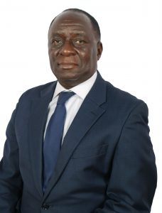
Michael Grace Kawooya is a Professor of Radiology at the Ernest Cook Ultrasound Research and Education Institute and Professor Emeritus at the Makerere University College of Health Sciences in Kampala, Uganda. He has done much for the development of radiology in his country and the rest of Africa, but says efforts must continue to increase the number of radiologists and range of equipment, and to raise awareness of radiation safety. Kawooya believes Africa can learn a lot from European advances. His contributions to improving bilateral cooperation will be rewarded today as he receives ESR Honorary Membership.

Professor Michael G. Kawooya’s contributions to improving bilateral cooperation between Africa and Europe will be rewarded today as he receives ESR Honorary Membership.

ECR Today: How much has radiology advanced in Uganda?

Michael Grace Kawooya: Back in the late 1980s, radiology was very new to medical practice in Uganda, and its contribution to healthcare was not well understood. It was not given priority and was underfunded. There were only two radiologists in the country when I finished my residency and we were overwhelmed with work. Doctors were not willing to undertake radiology residency, fearing that radiologists didn’t earn much. Equipment was scanty and often malfunctioning. Many of these challenges still exist today, but to a lesser extent. Radiology is better understood and its role is now evident. The number or radiologists has increased to almost 70. This year alone, 20 doctors took up radiology residency. The number and range of equipment has also increased.

ECRT: Are there any regional trends in radiology in Africa?

MGK: The same challenges facing radiology in Uganda bedevil most of Africa, but North Africa, which is largely Arabic, and South Africa, which is wealthier, face fewer challenges. In these parts of Africa, radiology has flourished more compared to Central, East, and West Africa. The radiologist-to-population ratio is approximately 1:67,000 in Egypt, 1:1,600,000 in Uganda and 1: 8,000,000 in Malawi. The more affluent regions have higher numbers and range as well as sophistication of imaging equipment. They have more radiology training institutions and undertake more research. Read more…

Ex-RSNA chief calls for more international cooperation

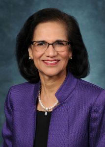
Vijay M. Rao, MD, FACR is the David C. Levin Professor and Chair of the department of radiology at Thomas Jefferson University. She is also senior vice-president of Enterprise Radiology, Jefferson Health, and the immediate past president of the Radiological Society of North America. As she receives ESR Honorary Membership today, she looks back on 30 years of work in a field she describes as globally united.

Radiology has come a long way from the days of plain film hypocycloidal tomography in head and neck imaging, a subspecialty to which Prof. Rao has dedicated most of her career. Technological innovations such as ultrasound, CT, MRI and PET have revolutionised the field, allowing for earlier diagnosis and greater insights into the pathophysiology of a host of diseases and conditions.

Professor Vijay M. Rao from Philadelphia, immediate past president of the RSNA, will receive ESR Honorary Membership today during the Grand Opening.

Radiology continues to evolve from anatomic to more physiologic and functional assessment of disease processes, with more precise quantification and minimally invasive therapeutic options.

Rapid technological advances have led to tremendous growth in radiology along with associated costs. But the digital age has introduced a number of previously non-existent opportunities to transform radiology practice, using tools powered by informatics and machine learning (ML). “Radiologists should explore ways to utilise these technological advances to add value and reduce waste in healthcare,” she said.

Artificial intelligence (AI) and ML applications are valuable tools that make radiologists more effective and increase their contributions to personalised and precision medicine. “AI can assist radiologists at a time when we are challenged to provide imaging services that are faster, safer and affordable, as well as information that is quantitative and precise. These new technologies will improve workflows in our daily practice, freeing up time for us to better position ourselves as integral members of the patient’s healthcare team,” she said.

The move toward value-based imaging continues to be the biggest trend in the US, where emphasis is placed on volume when structuring radiology practice. “Policymakers lack understanding of imaging’s contribution to patient care and the extent of services that radiologists provide. That is why we have begun the necessary transition to a value-based model of care, where radiologists are actively consulting with patients and their referring physicians as part of the healthcare team,” she explained. Read more…

Keep ultrasound close and generate added value, Lubinus advises

Professor Federico G. Lubinus is full professor of radiology and director of the radiology residency programme at the autonomous university of Bucaramanga (UNAB) in Colombia. He is one of the leaders in South American radiology and he believes that bringing back ultrasound to the heart of radiological practice is key to the future growth of the specialty. His efforts will be acknowledged today as he receives ESR Honorary Membership.

When it comes to radiology, South America has very similar challenges to the rest of the world and a number of regional trends, Lubinus explained. “Big workloads, turf battles with other medical specialties, and challenges imposed by teleradiology and artificial intelligence are some of the many difficulties we have to deal with, not only as radiological societies but also as radiologists wondering about the role of the specialty in the near future,” he said.

Professor Federico G. Lubinus, who will receive ESR Honorary Membership at today’s Grand Opening, is one of the leaders in South American radiology.

But radiology practice in the continent is uneven and a major difference concerns radiology training, according to Lubinus, who currently heads the Latin American Educational Forum, a group that brings together radiological societies of South America to develop and advance educational issues in the region. “Training is dependent on the regulations of each country. There is great diversity both in the number of years of study required to be a specialist and in the minimum requirements demanded in each country, which makes it difficult to standardise programmes and perform appropriate evaluation of knowledge,” said Lubinus.

Lubinus is also director of the scientific committee of the Asociación Colombiana de Radiología (ACR), a society he served twice as president. Under his aegis, the ACR worked to have a standardised training programme, a tool that is now recognised by the Colombian ministry of education as the basis for evaluation and certification of radiology graduates, and radiologists who have received their certification abroad.

For the past four years, the Latin American Education Forum has also engaged in developing a standardised academic residency programme as a reference instrument for the validation of qualifications of Latin American radiologists, to improve the much needed certification and recertification in radiology.

Read more…

Russia’s preeminent radiologist continues to reach for the top, after two decades with the ESR

Professor Valentin Sinitsyn is chair of radiology and head of the radiology department at the medical faculty of Moscow Lomonosov State University, Moscow. A pioneer in cardiac imaging in Russia, he has helped advance the field in his country, but believes there is still much to be done to increase MR and CT use. After 20 years of working to advance the ECR and European radiology, he will receive the ESR Gold Medal today.

Sinitsyn has witnessed the introduction of spiral and multidetector CT, high-field MRI and hybrid imaging, and the development of evidence-based radiology and medicine.

Professor Valentin Sinitsyn from Moscow

Professor Valentin Sinitsyn from Moscow, a pioneer in cardiac imaging in Russia, will receive the ESR Gold Medal today.

As he graduated from Sechenov Medical University in Moscow in 1984, cardiac CT and MRI did not yet exist. “With the help of these new modalities, radiology penetrated fields where it had never been used before, and the best example is cardiac imaging,” he said.

Sinitsyn started implementing cardiac imaging back in the 1980s and has very fond memories from that time. Although these were also harsh days for the field, he said.

“Most cardiologists and radiologists believed that cardiac MRI and CT were toys that were good for nothing in real clinical practice. I still remember my first case of apical hypertrophic cardiomyopathy diagnosed with cardiac MRI in 1987, when nobody could make a correct diagnosis,” he said.

Today he admits to being a little disappointed with the slow penetration of cardiac MRI and CT in Russian clinical practice. The benefits of modern cardiac imaging are well known, but the percentage of cardiac CT and MR examinations in cardiac imaging is just around 1%. “Low reimbursement for such examinations and insufficient knowledge and motivation from both radiologists and cardiologists are mostly to blame for this situation,” he said.

Read more…

Trojanowska to deliver honorary lecture on neglected form of cancer

Polish radiologist Dr. Agnieszka Trojanowska will shed light on human papilloma virus (HPV)-induced squamous cell cancer of the head and neck, a common yet long neglected type of cancer. In an interview with ECR Today, she also explained the origins of her passion for head and neck imaging, and how radiologists will increasingly benefit from computer science.

Dr. Agnieszka Trojanowska, assistant professor in the department of radiology and nuclear medicine at Lublin University Hospital, is an internationally recognised specialist in head and neck imaging. Her special interest for oncology and her natural curiosity led her to agree to deliver the Josef Lissner Honorary Lecture on HPV-induced squamous cell cancer of the head and neck (HNSCC) at ECR 2018.

Agnieszka Trojanowska from Lublin, Poland, will speak on HPV-induced squamous cell cancer of the head and neck in her Honorary Lecture today.

“HNSCC behaves differently than more well-known forms of SCC, and it is becoming more and more prevalent. It affects young, well-educated people with high socioeconomic status, and is a quickly rising sexually transmitted entity with peculiar clinical and molecular characteristics. In particular the rise of HPV-induced cancer has been observed in the United States since the beginning of 21st century,” she said.

It was important for her to talk about this long overlooked topic, for which much remains to be done. “It has been under-estimated for many years in many countries. Nowadays, we can speak about an HPV epidemic, leading to a significant rise of oropharyngeal cancer incidences worldwide. These cancers are quite unique and, in my opinion, knowledge of their physiology and treatment options is essential,” she said.

New research shows that, compared with environmental-related head and neck squamous cell carcinomas, patients with HPV-related malignancies display a better response to treatment and a lower risk of death and progression, Dr. Trojanowska pointed out. It is also noteworthy that, according to the 2017 TNM classification, HPV+ cancers are evaluated with a separate grading scale, she added.

Read more…