ECR continues to think outside the box and adds the Cube

For young physicians, interventional radiology (IR) may seem as enigmatic as a Rubik’s cube. But the cube can be solved with the right algorithm, according to Dr. Maximilian de Bucourt, head of angiography at Charité Campus Benjamin Franklin in Berlin.

“What we do in IR is exactly like a Rubik’s cube: you follow the algorithm and solve the problem. In IR, if you do the procedure steps over and over again and use the rules, most of the time you can get the solution for the patient,” he said.

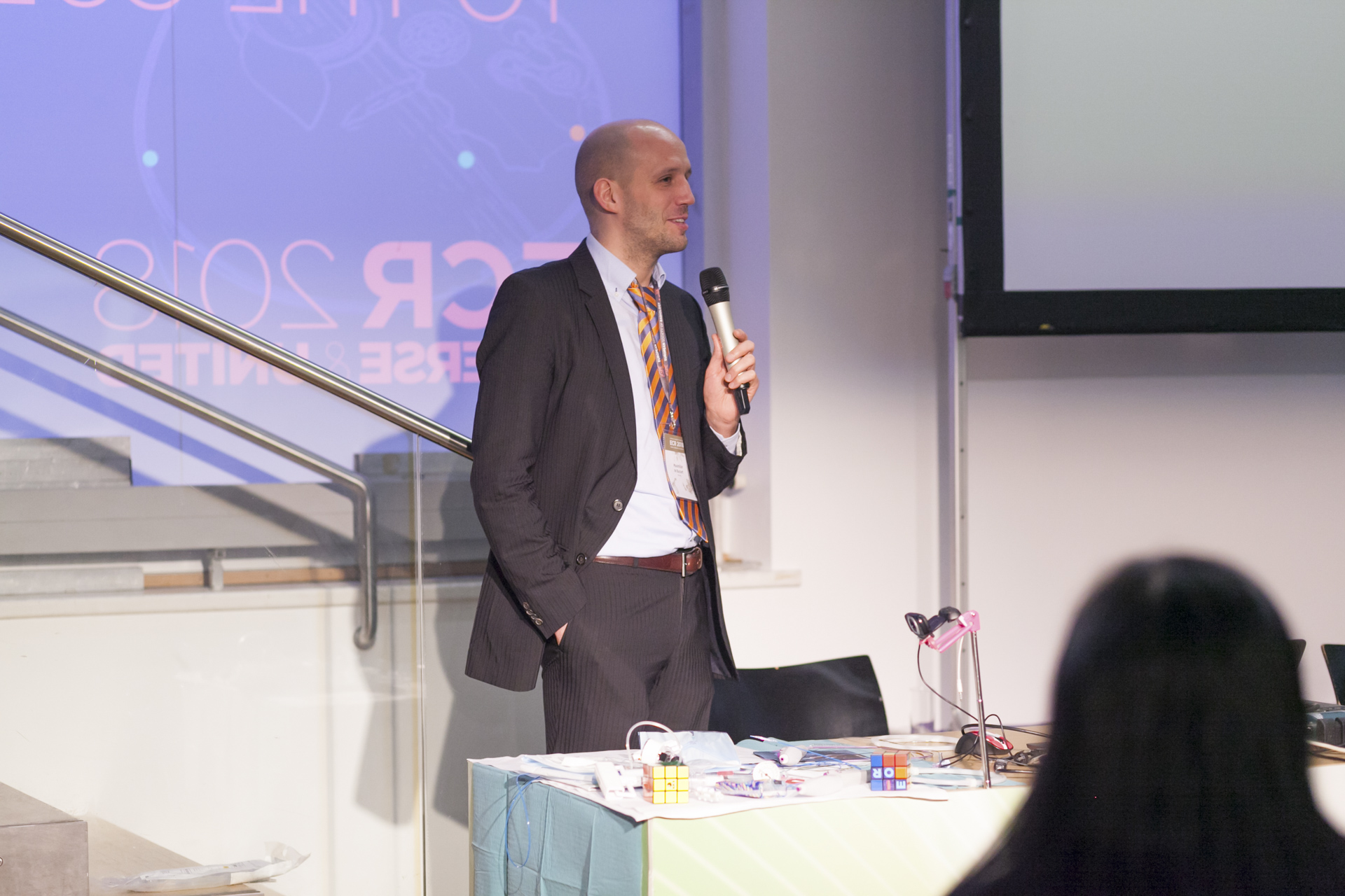
Dr. Maximilian de Bucourt is head of angiography at Charité Campus Benjamin Franklin in Berlin, and one of the specialists behind the Cube.

Together with Prof. Christian Loewe from Vienna, de Bucourt imagined ‘the Cube’, a workshop aimed at introducing young physicians to the tools and techniques used in IR. With its focus on hands-on activities, including simulated procedures and interactive demonstrations, the Cube is fulfilling its goal of acquainting residents with this unique sub-specialty.

“The Cube is for young people who are thinking of becoming IR specialists, but find it too long before they can deploy a stent or manoeuver a catheter inside an artery. After medical school, basic radiology training and dedicated IR training with a teacher willing to let them perform major interventions, they still need to learn how to manage complications. The Cube is all about expediting this process, by enabling students and residents to get their hands on the products earlier,” he explained. Read more…

Riklund to unveil the next big thing in hybrid imaging

The Swedish radiologist Prof. Katrine Riklund, the current Chair of the ESR Board of Directors, has dedicated her career to the development of hybrid imaging. During todays’ Marie Curie Honorary Lecture (Room A, 1:00 p.m.) she will look back at the achievements made in this emerging field and look forward to future advances.

Riklund, who is a professor, consultant in diagnostic radiology and pro-vice-chancellor of Umeå University, is one of Sweden’s leading radiologists. She is also one of the first researchers to have recognised the potential of combining PET with CT and MRI, and has worked to advance the field ever since its emergence at the beginning of the 21st century.

“The combination of structural and functional/molecular imaging is fascinating. The entire field is new and extremely interesting, and it’s the closest to my heart when it comes to imaging,” she said.

Prof. Katrine Riklund from Umeå, Sweden, will speak about the growing significance of hybrid imaging in today’s honorary lecture.

During her lecture, Prof. Riklund will share examples of what hybrid imaging can do and what is going to come next. A major trend will be making use of the entire hybrid imaging examination for diagnostic protocols, also for the CT part, she believes. “This changes workflow and gives us more information. PET and CT or MR are not competing techniques, they are complementary,” she said.

Tracer development is key for PET but digital detectors will also represent a major step forward. Currently, the need for a cyclotron for production of radionuclides hinders substantial distribution of scanners outside large centres. “To make hybrid imaging really take off, we need other forms of tracer production. I would like to see the tracer production work like a coffee machine – with buttons to select tracer and radionuclide,” she said.

In her day-to-day work, Prof. Riklund is involved in various research projects, such as COBRA, a prospective multimodal imaging study of dopamine, brain structure and function, and cognition; (PEARL-PD), 18F-FE-PE2I PET/CT, a study of dopamine transporters in early Parkinson’s disease, RECTOPET (REctal Cancer Trial On PET-MR/CT); and Prostate Cancer – PSMA and Acetate in PET/CT and PET/MR. These projects reflect her three major interests in the field; movement disorders and cognition in central nervous system, and prostate and colorectal cancer.

Read more…

The future is now, Marc Dewey says

The radiological community must understand the potential of value-based radiology and its related challenges, the German radiologist Marc Dewey will argue during the Wilhelm Conrad Röntgen honorary lecture today at ECR 2018.

Marc Dewey is Heisenberg Professor of Radiology of the German Research Foundation and Vice Chair of the Department of Radiology at Charité – Universitätsmedizin Berlin. His main research interest is in cardiovascular imaging but he is interested in many other aspects of clinical practice including value-based imaging, something he will detail in his lecture at the ECR.

Prof. Marc Dewey from Berlin will deliver today’s Honorary Lecture on value-based imaging at 12:15 in Room A.

“Participants will get to know three things. First, why the time for value-based radiology is now. Second, they will appreciate the potential of value-based radiology in the clinical care process. And third, understand the challenges in implementing value-based radiology,” Dewey promised.

Combining human image analysis and artificial intelligence has great potential for creating value for patients at lower costs, he believes. “This is value-based radiology and the time to get involved is now, as this is not merely a new technical toy of radiologists but will be accompanying the entire diagnostic and treatment pathway in all clinical service lines for the benefit of our patients,” he said.

Dewey is the coordinator of the DISCHARGE trial, an EU-funded multicentre project with more than 30 partners across Europe that will determine whether cardiac CT may replace invasive coronary angiography in certain patients. “The DISCHARGE project is a unique and truly impressive effort of several hundred individuals working at 31 sites in 18 European countries. External advisory board members were often thrilled when first witnessing the team spirit at our annual meetings,” said Dewey, who will soon present results of the on-going clinical trial. He said coordinating a large project on imaging in Europe gave him “the ability to better understand the culture, concerns, and ideas in different regions of Europe.”

Read more…

Facial genetics and forensics take centre stage at ECR

Watch this session on ECR Live: Friday, March 3, 10:30–12:00, Room B
#ECR2017

Forensics and facial genetics will be in the spotlight at ECR 2017 as the ‘ESR meets Belgium’ session offer a look at medical imaging’s most original contributions to healthcare and crime investigation.

Advanced decomposition of brain MRI or facial 3D surface images into modules, tailored for associations with underlying genetic variations.

Dr. Peter Claes from Leuven, Belgium, will present his work in facial genetics in a session titled ‘Imaging genetics and beyond: facial reconstruction and identification’.

Claes is a senior research expert in the Medical Image Computing research group of the Processing of Speech and Images division of the Electrical Engineering department at Leuven Catholic University.

He uses CT, MRI and 3D surface imaging modalities to grasp the link between people’s appearance and underlying genetic variations.

“Your appearance is genetically driven. In families there’s a strong link, even more so between identical twins, who share the same DNA profile and almost the same face. Physical features also influence your brain, the way you think. A lot of facial characteristics are shared, for example in Down’s syndrome patients, who present with the same features whether they are European or Asian,” he explained.

The link between genetic disorders and facial genes has been of interest to scientists for a while, but research is slow and tedious.

Claes became interested in facial genetics after working in craniofacial morphometrics to help correct morphological abnormalities and anomalies, and in craniofacial reconstruction to identify victims.

To help decipher facial genetics, Claes uses the computer-based craniofacial reconstruction programme he developed for victim identification, and combines 3D surface processing, statistical modelling, analysis, mapping and prediction techniques. He has also created an array of algorithms and software for investigators who plan to use 3D facial datasets. Last year, he also co-organised the first international workshop on facial genetics in London.

Read more…

Education central to improving imaging data quality in oncology clinical trials

Watch this session on ECR Live: Thursday, March 2, 16:00–17:30, Room X
#ECR2017

Imaging data is key in multicentre clinical trials for cancer research but quality control is currently a major impediment, bringing the validity of the trials into question and potentially impacting on the quality of drugs put on the market, a panel of experts will argue today in a session held by the ESR and the European Organisation for Research and Treatment of Cancer (EORTC) at the ECR.

Imaging is increasingly contributing to cancer research thanks to the development of innovative techniques that depict functional and molecular processes. In most oncological clinical trials, imaging is now the primary criteria used to evaluate progression of disease or efficiency of the drug being tested.

The best way to obtain valuable imaging measurements is to involve the imagers who take part in these trials and educate the clinician investigators, experts will explain in the session.

When it comes to imaging in cancer research, a number of issues take centre stage. Difficulties associated with integrating imaging biomarkers into trials have been neglected compared with those relating to the inclusion of tissue and blood biomarkers, largely because of the complexity of imaging technologies, safety issues related to new contrast media, standardisation of image acquisition across multivendor platforms and various post-processing options available with advanced software, as reported recently in The Lancet by the EORTC and leading researchers.

Read more…

01
Mar 2017
POSTED BY
POSTED IN ,
DISCUSSION 0 Comments

Eminent neuroradiologist to give honorary lecture at ECR 2017

Watch this session on ECR Live: Thursday, March 2, 12:15–12:45, Room A
#ECR2017

In recognition of his significant impact on the field of neuroimaging and his service to organised radiology, Professor Mauricio Castillo from Chapel Hill, NC, United States, has been invited to deliver the Wilhelm Conrad Röntgen Honorary Lecture ‘Dissatisfaction, burnout and inequality: three major challenges in radiology’ at ECR 2017.

Professor Mauricio Castillo from Chapel Hill, United States

Professor Mauricio Castillo from Chapel Hill, United States, will deliver the Wilhelm Conrad Röntgen Honorary Lecture today at 12:15 in Room A.

Mauricio Castillo is the James H. Scatliff distinguished professor of radiology, chief and programme director of neuroradiology at the University of North Carolina in Chapel Hill. He is currently president of the American Roentgen Ray Society.

Originally from Guatemala, Prof. Castillo completed his radiology and neuroradiology training at the University of Miami School of Medicine, Jackson Memorial Medical Center, and Emory University School of Medicine, Affiliated Hospitals in Atlanta respectively.

His specialty and subspecialty choice was in large part motivated by his mentors.

“When I was a medical student, I was fortunate to work in the only teaching hospital that had a CT scanner and two board-certified radiologists. I was very impressed by the impact of imaging in patient care and by the way these two professionals used their large base of knowledge of medicine to generate a differential diagnosis based on imaging findings. Later on, while I was a resident, I developed a close relationship with Dr. Robert Quencer, which led me to become a neuroradiologist. One should never underestimate the influence we may have on our trainees and the influence our mentors have had on us,” he said.

Prof. Castillo’s research interests include paediatric neuroimaging, application of new imaging techniques and medical literature editing. Read more…

Prominent Brazilian radiologist to shed light on Zika virus imaging at ECR 2017

In recognition of her major achievements in neuroimaging and advancement of the field, Professor Maria de Fátima Viana Vasco Aragão from Recife, Pernambuco, Brazil, will present the Guest Lecture ‘Breaking News from Latin America: How to recognise Zika virus infections on imaging studies’ at ECR 2017.

Maria de Fátima Viana Vasco Aragão is professor of radiology at the Maurício de Nassau University and Scientific Director of the Multimagem Diagnostic Centre in Recife, Brazil. She is also financial director of the Diagnostika Endoscopy and Radiology Clinic in Recife.

Prof. Vasco Aragão received her medical degree from Pernambuco Federal University in 1987 and completed her residency in radiology at Ribeirão Preto Faculty of Medicine, University of São Paulo. She did fellowships in CT and MRI and in neuroradiology at the Med Imagem Beneficencia Portuguesa, São Paulo, and later completed a research fellowship at Mount Sinai Hospital Center in New York, US.

She always knew she would become a doctor and chose radiology because she recognised early on the field’s value in diagnosis and treatment.

Maria de Fátima Viana Vasco Aragão is professor of radiology at the Maurício de Nassau University and Scientific Director of the Multimagem Diagnostic Centre in Recife, Brazil.

Maria de Fátima Viana Vasco Aragão is professor of radiology at the Maurício de Nassau University and Scientific Director of the Multimagem Diagnostic Centre in Recife, Brazil.

“My mother is a retired biologist and biology teacher. I have never forgotten the moment she taught me what a cell was. In my family, educators predominated and my great aunt was the first woman in Pernambuco, my home state, to graduate from medical school, in 1935. Perhaps on account of this family background I made up my mind to become a doctor. As an intern student of medicine, I was very impressed when I took my patients to be examined by CT and US. Even though there was only one CT scanner in my city, I succeeded in having all my intern patients examined, when indicated. My tutors were impressed by the swiftness with which cases were resolved. Thus I learned that radiology not only helps to quickly define diagnosis in most patients, but also provides a clear prognosis for the most adequate treatment. At the end of the course I began considering the possibility of specialising in radiology, even though 29 years ago, for most people, a radiologist was not a doctor. Shortly after graduation, I sat for a test and won a place in radiology at the first attempt, and radiology has been my passion ever since. I would like to say that I am grateful for having had great mentors, including the neuroradiologist Dr. Sérgio Santos Lima, who was my director during my fellowship in the Med Imagem Beneficencia Portuguesa, and the head and neck radiologist, Dr. Peter Som, who was one of my directors during my research fellowship at Mount Sinai Hospital Center New York,” she said.

Read more…

Hybrid imaging contents to heighten delegates’ interest at ECR 2016

ECR2015_ECRToday_Blog

ECR Today spoke with ECR 2016 Congress President, Prof. Katrine Åhlström Riklund, deputy head of the department of radiation sciences and director of the medical school at Umeå University, Sweden, to find out a little bit about next year’s annual meeting.

ECR 2016 Congress President, Prof. Katrine Åhlström Riklund, from Umeå, Sweden.

ECR 2016 Congress President, Prof. Katrine Åhlström Riklund, from Umeå, Sweden.

ECR Today: What will be the highlights of ECR 2016?
Katrine Åhlström Riklund: It is hard to tell what the specific highlights will be more than one year ahead, due to the rapid development of imaging. The highlights will be the entire congress through its well-developed programme, which covers the whole range of education from student level to advanced subspecialists. I should say the added content of hybrid imaging in several sessions would make the programme even more attractive. Besides the educational and scientific programme, the grand opening ceremony and social activities will also be memorable events.

ECRT: Will there be any new additions to the programme?
KAR: As always, there will be innovations at the ECR. The content of hybrid imaging will be spread across several sessions and not in one single session. The new session formats introduced at ECR 2015, with the European Excellence in Education (E3) programme – divided into five levels (the Rising Stars programme, European Diploma Prep Sessions and Beauty of Basic Knowledge programme, ECR Academies and ECR Master Classes) will be continued. These levels cover the entire span from undergraduate medical education to subspecialised continuing professional development. Getting involved in the sessions is important for retaining knowledge.

Read more…

Comprehensive personalised imaging transforms cardiothoracic disease management

ECR2015_ECRToday_Blog

Watch this session on ECR Live: Sunday, March 8, 8:30–10:00, Room E1
Tweet #ECR2015E1 #NH17

Besides personalised imaging, a new paradigm is emerging in radiology that should re-shape clinical practice and benefit the patient immensely. Supported by new technologies that enable radiologists to image the body faster and better, radiologists are now trying to broaden their focus during examinations.

If there is a field where these advances make a tremendous difference, it is cardiothoracic imaging, an area where diseases are more often than not intertwined. Cardiovascular and chest radiologists will explain how the comprehensive personalised approach impacts their work and try to convince radiologists on both sides to take an interest in the other, in a New Horizons session on Sunday at the ECR.

For years, the trend was for radiologists to subspecialise as much as they could. Cardiovascular radiologists and chest radiologists would focus on their own area with little or no interest beyond that. But among these subspecialists, an increasing number are now changing their approach, as mounting evidence shows that diseases of the heart and chest are very often related, according to Dr. Christian Loewe, deputy head of the section of cardiovascular and interventional radiology at the Medical University of Vienna, Austria.

Dr. Christian Loewe is deputy head of the cardiovascular and interventional radiology section at the Medical University of Vienna, Austria.

Dr. Christian Loewe is deputy head of the cardiovascular and interventional radiology section at the Medical University of Vienna, Austria.

“In the past patients were investigated by either focusing on chest or cardiac diseases. This choice was mainly driven by their first clinical examination. However, there are a lot of situations and diseases where chest problems are caused by cardiac diseases and vice versa. There’s a huge interaction between heart and chest, and that’s why it’s interesting and important to look at this relationship in more detail today,” he said.

To prove his point, Loewe, a cardiovascular radiologist, will talk about acute and chronic chest pain during the session. Some of the most severe causes of chest pain are due to cardiovascular diseases, including myocardial infarction or acute aortic diseases. However, acute chest pain can also be caused by a number of pulmonary diseases, including pulmonary embolism, pneumonia and others. Therefore, radiologists must learn the different life-threatening disorders that cause chest pain, whether they are respiratory or cardiovascular.

Read more…

Population imaging studies gain ground in healthcare

ECR2015_ECRToday_Blog

Watch this session on ECR Live: Thursday, March 5, 16:00–17:30, Room L1
Tweet #ECR2015L1 #PC8B

Imaging large cohorts of people enables scientists to collect information useful for science and emphasises radiology’s role in healthcare. From the most recently available imaging biomarkers to data such as genomics and metabolomics, Thursday’s dedicated Professional Challenges Session will show just how useful population imaging studies have become in the prognosis of countless diseases.

When radiologists follow a cohort of people for 20 or 30 years, the benefits for the patient increase tremendously. If a radiologist performs a CT examination of a patient’s coronary arteries and finds calcification, chances are that the patient will have a heart attack within the next few years. Unfortunately at this stage, the patient is usually out of the radiologist’s reach.

The fluid-attenuated inversion recovery axial image of a 26-year-old female participant shows multiple, bilateral, asymmetric, linear and ovoid hyperintensities that are located perpendicular callososeptal characteristic of multiple sclerosis

The fluid-attenuated inversion recovery axial image of a 26-year-old female participant shows multiple, bilateral, asymmetric, linear and ovoid hyperintensities that are located perpendicular callososeptal characteristic of multiple sclerosis

However, if patients chose to participate in a population study, they will be checked on a regular basis, and radiologists will be able to access previous information and initiate appropriate treatment earlier, significantly
improving patient outcome.

Securing imaging data is always tricky and population imaging studies are an opportunity for radiologists to access this data. Showing the relevance of imaging findings highlights radiology’s role in the medical continuum, according to Prof. Norbert Hosten, of the Ernst-Moritz-Arndt University in Greifswald, Germany, who will chair
the session Thursday.

“Our way to prove that radiology can make people healthier and happier is to do large population imaging studies. Radiology can develop the kind of data that are necessary to prove that our methods really help the patient,” he said.

Read more…