B-0889 Improved image quality for higher diagnostic accuracy of cranial computed tomography using iterative image reconstruction
H. Haubenreisser, C. Fink, P. Apfaltrer, B. Schmidt, M. Sedlmair, S.O. Schönberg, T. Henzler | Monday, March 11, 14:00 – 15:30 / Room B
Purpose: To prospectively compare the image quality of cranial computed tomography (cCT) with thin slice widths using traditional filtered back projection (FBP) and sinogram-affirmed iterative image reconstruction (SAFIRE).
Materials and Methods: 40 consecutive studies (19 men; 71.6±16.6 years) referred for cCT were prospectively included. Each cranial CT raw data set was reconstructed with FBP and SAFIRE with decreasing slice widths (5 mm-1 mm). Objective image quality was assessed by measuring image noise in three predefined regions of the brain (white matter, thalamus, cerebellum) using identical regions of interest (ROIs). Subjective image quality was assessed by 2 experienced radiologists by ranking the reconstructed data sets with respect to overall image quality. The Mann-Whitney U-test and Cohen’s Kappa were used for statistical analysis.
Results: Image noise was statistically significantly reduced in all SAFIRE images at identical slice widths when compared to the images reconstructed with FBP (4.26±0.43 HU vs. 7.67±1.19 HU at 1 mm slice width) (p<0.001). Mean signal attenuation for each region and slice width remained constant between the two reconstruction methods (p>0.5). SNR was comparable between 1 mm SAFIRE images and 5 mm FBP images. Subjective image quality of SAFIRE images was rated consistently higher than that of the FBP images (p<0.001). Interobserver agreement was excellent between both radiologists (Cohen’s K = 0.79-0.86).
Conclusion: Iterative image reconstruction significantly reduces image noise, while increasing image quality. In cCT this may be used to decrease slice width and thus reduce partial volume effects, which may lead to increased diagnostic accuracy.

Dear Friends,
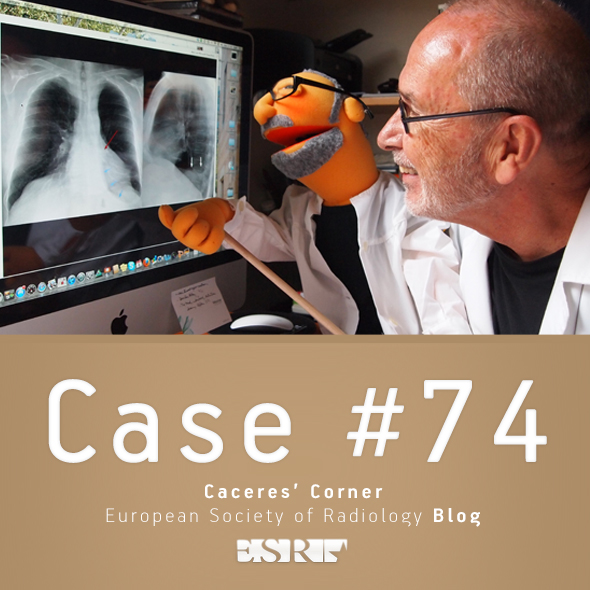
This year I plan to show only chest cases, emphasising the diagnostic approach to basic patterns in the plain film. Hopefully, this monographic approach will help you with the diploma examination.
Cases will be posted every other Monday and answers will be given on Friday.
Radiographs (below) of the first case belong to a 57-year-old man, asymptomatic. Study them carefully, leave your opinions in the comments section, and look out for the answer on Friday.
Diagnosis:
1. Thymoma
2. Teratoma
3. Mediastinal fat
4. Can’t tell
Read more…

A-093 Cardiomyopathies
P. Sipola | Friday, March 8, 08:30 – 10:00 / Room L/M
Cardiac magnetic resonance imaging (CMRI) is highly valuable in the differential diagnosis of cardiomyopathies. MRI diagnosis is based on cine imaging of cardiac function, T2-weighted imaging of oedema and late gadolinium-enhanced (LGE) patterns of scar tissue. Hypertrophic cardiomyopathy (HCM). Left ventricular hypertrophy (LVH) is typically located in basal septum and anterior wall but has variable expression (diffuse, localized). Associated abnormalities include left ventricular (LV) high-ejection fraction (EF), mitral valve abnormalities, apical aneurysm, and right ventricular (RV) hypertrophy. Scattered intramyocardial LGE may occur in various patterns. The differential diagnoses in patient with hypertrophic phenotype include pressure overload hypertrophy, amyloidosis, sarcoidosis, and Fabry’s disease. LGE patterns is useful in differentiation. Dilated cardiomyopathy (DCM): Dilated LV end-systolic volume and impaired EF% are characteristics. Non-ischaemic DCM typically shows no LGE (in contrast to ischaemic cardiomyopthy). Sometimes faint midwall enhancement can be observed, which has prognostic value. Presence of extensive non-compacted myocardium indicates non-compaction cardiomyopathy. Arrhythmogenic right ventricular cardiomyopathy (ARVC): The RV volume is enlargened and akinetic RV segments can be seen. Local bulging or dyskinesia in conjunction with fatty infiltration and LGE is typical. Restricted cardiomyopathy (RCM): Enlargened atrias and normal sized ventricles with preserved EF% and no LGE are characteristics. Myocarditis: LV systolic function is typically lowered but may be normal. T2 images may show increased signal. LGE limited to the subepicardial myocardium is highly suggestive of myocarditis. Iron overload cardiomyopathy: Cine imaging is used to assess LV global function and T2*-weighted imaging to quantitate ventricular iron deposition.

Dear friends,
After a two-month vacation, we return with renewed energy. I had a long conversation with Dr. Pepe and we decided to present our cases alternately: I will show my cases one week and Dr. Pepe will present the diploma cases the following week. Cases will be posted on Monday morning and answers will be offered on Friday. This way we will not compete with each other and can remain good friends.
Considering that most of you are still on vacation or with a half-functioning brain, Muppet has selected an easy warm-up case of a 46-year-old woman, asymptomatic.
As usual, leave your thoughts and answers in the comments section below.
Diagnosis:
1. Filariasis
2. Talc aspiration
3. Cysticercosis
4. None of the above
Read more…

B-0688 One-to-one comparison between digital mammography and digital breast tomosynthesis using a fully automated software: breast density underestimation on digital breast tomosynthesis varies in different BI-RADS classes
A. Tagliafico, S. Airaldi, F. Cavagnetto, B. Bingotti, S. Tosto, D. Astengo, M. Calabrese | Sunday, March 10, 10:30 – 12:00 / Room F2
Purpose: To compare breast density on digital mammography (FFDM) and tomosynthesis (DBT) according to different BI-RADS classes (four classes from 1 to 4) with an automated software.
Methods and Materials: IRB approval and written informed consent were obtained. Digital breast tomosynthesis and digital mammography were obtained in the same patient. A total of 160 consecutive patients (mean age years: 50±14; mean BMI: 22 ± 3) were included. One-to-one comparison between FFDM and DBT was made with a fully automated software previously validated. Statistical analysis was performed with two-tailed t-test for paired data using statistical software.
Results: In BI-RADS class 1, digital mammography overestimated breast density of a 16 %. In BI-RADS class 2, digital mammography overestimated breast density of a 11.9%. In BI-RADS class 3, digital mammography overestimated breast density of a 3.5%. In BI-RADS class 4, digital mammography overestimated breast density of a 18.1%. The differences resulted highly statistically significant (p<0.0001). There was a good correlation between BI-RADS categories and the density evaluated with digital mammography and digital breast tomosynthesis (r=0.56, p <0.01 and r=0.48 p<0.01).
Conclusion: Breast density values were underestimated by DBT in comparison to FFDM with a non-linear relationship in the different BI-RADS classes. This data should influence clinical and research studies dealing with breast density as a qualitative biomarker.

B-0984 Hepatic parenchymal and vascular contrast improvement in super-delayed phase images of Gd-EOB-DTPA-enhanced MRI
S. Kobayashi, O. Matsui, T. Gabata, W. Koda, T. Minami, K. Kozaka, A. Kitao | Monday, March 11, 14:00 – 15:30 / Room I/K
Purpose: To elucidate the parenchymal and vascular contrast improvement effect of super-delayed phase (SDP) images of Gd-EOB-DTPA (EOB)-enhanced MRI in poor hepatobiliary phase (HBP) image cases special focus on Child-Pugh (CP) classification.
Methods and Materials: 76 cases, who have examined EOB-enhanced MRI for closer examination of hepatic lesions, and taken SDP images approximately 90 minutes after iv administration of EOB because of poor HBP image are subjected to this study. 20 hepatobiliary disease cases who had also taken SDP images which show normal HBP images were used as control. Hepatic vascular/parenchymal enhancement ratios (ER) were defined as signal intensity (SI) of intrahepatic vessel / SI of liver. ER of HBP and SDP were calculated and compared between each CP class liver damage groups. Chi square test was used for statistics and p<0.05 was considered statistical significant.
Results: In poor HBP cases (n=76), ER of HBP and SDP were 0.88±0.16 and 0.64±0.16. In control cases (n=20), ER of HBP and SDP were 0.54±0.08 and 0.39±0.06. ER of HBP and SDP in CP-A poor HBP (n=27), CP-B poor HBP (n=47), CP-C poor HBP (n=2) were 0.83±0.14 and 0.60±0.13, 0.90±0.16 and 0.65±0.16, 1.03±0.16 and 0.99±0.19, respectively (all combinations except CP-C showed significance difference).
Conclusion: In most of the poor HBP image cases, SDP image improve parenchymal and vascular contrast except CP-C liver damage cases.

B-0789 CT colonography: accurate registration of prone and supine endoluminal surfaces of the colon
T.E. Hampshire, H.R. Roth, E. Helbren, A. Plumb, D. Boone, G. Slabaugh, S. Halligan, D.J. Hawkes | Monday, March 11, 10:30 – 12:00 / Room E2
Purpose: Computed tomographic (CT) colonography is a technique for detecting bowel cancer or potentially precancerous polyps. Because retained fluid and stool can mimic pathology, CT data are acquired with the patient in both prone and supine positions. Radiologists then match endoluminal locations between the two acquisitions to determine whether pathology is real. This process is hindered by the fact that the colon can undergo large deformations that often occur during repositioning of the patient. Automated registration between datasets could potentially improve efficiency and diagnostic accuracy.
Methods and Materials: We have developed software to establish correspondence between prone and supine endoluminal surfaces. An initialisation step generates image patches at the positions of haustral folds using depth map renderings and is optimised by virtual camera registration. Additional neighbourhood information is then included in a Markov Random Field model to establish landmark-based correspondences. Subsequently, the complexity of the registration task is reduced by mapping both prone and supine surfaces onto a cylindrical domain in which correspondence is established using non-rigid image registration.
Results: The registration was applied to 17 CTC cases including cases exhibiting luminal collapse, achieving fold matching accuracy of 96 %. Providing an accurate initialisation, the method significantly improved the cylindrical registration (p<0.001), achieving a mean error of 6.0mm measured at 1743 reference points.
Conclusion: The proposed method can successfully establish correspondence between prone-supine locations on the endoluminal surface derived from CT colonography. The ability to rapidly and automatically match polyps between acquisitions will facilitate CT colonography interpretation.

B-0680 Texture analysis of malignant breast tumours: is a differentiation of ductal carcinoma in situ, invasive ductal and invasive lobular breast cancer possible?
T. Knogler, K. Pinker-Domenig, N. Perry, S. Milner, K. Mokbel, M.E. Mayerhoefer | Sunday, March 10, 10:30 – 12:00 / Room F2
Purpose: To evaluate the ability of texture features (TF), to differentiate between ductal carcinoma in situ (DCIS), invasive ductal carcinoma (IDC) and invasive lobular carcinoma (ILC) of the breast on full-field digital mammograms (FFDM).
Methods and Materials: 110 screen detected and histopathologically verified breast cancers (27 DCIS, 73 IDC, 10 ILC) imaged with FFDM in standard views were included in this study. For each lesion, a region of interest (ROI) was manually defined, which covered the lesion as well as a rim (1cm width) of normal-appearing breast tissue around the lesion in the view, where the lesion was depicted in largest diameter. TF derived from the grey-level histogram, co-occurrence matrix (COC), run-length matrix (RLM), absolute gradient (AG), autoregressive model (ARM) and wavelet transform were calculated for the ROIs. Fisher coefficients were calculated to determine which TF were best-suited for distinguishing between DCIS, IDC and ILC. Lesion classification was performed using linear discriminant analysis in conjunction with a k-nearest neighbour classifier, based on the combination of the 10 TF with the highest Fisher coefficients. Classification accuracy was used as the primary outcome measure.
Results: The accuracy of texture-based lesion classification was 84.33% (70 of 83 lesions) for IDC vs. ILC, 81.1% (30 of 37 lesions) for ILC vs. DCIS, but only of 70 % (70 of 100 lesions) for IDC vs. DCIS.
Conclusion: TF derived from FFDM may be of value for differentiating between ILC and IDC, and ILC and DCIS, but of limited value for differentiating between IDC and DCIS.

A-162 Upper limb nerve entrapment
D. Weishaupt | Friday, March 8, 16:00 – 17:30 / Room E1
The peripheral nerves of the upper limb are affected by a number of entrapment and compression neuropathies. These syndromes involve the brachial plexus as well as the musculocutaneous, axillary, suprascapular, ulnar, radial and median nerves. Clinical examination and electrophysiological studies are traditionally the mainstay of diagnostic work-up. However, ultrasonography and magentic resononance imaging (MRI) may provide key information about the exact anatomic location of the lesion or may help to narrow the differential diagnosis. In certain patients with the diagnosis of a peripheral neuropathy, imaging using either ultrasononography of MRI may help establish the cause of the condition and provide information crucial for conservative management or surgical planning. In addition, imaging is particularly valuable in compex cases with discrepant nerve functions test results.

A-495 Imaging of the most frequent emergencies of the genitourinary tract
L.E. Derchi | Sunday, March 10, 16:00 – 17:30 / Room E1
This presentation will deal with three of the most common and important acute problems of the GU system: testicular and ovarian torsion and the renal colic. US is the technique of choice in patients with acute scrotum and is able to identify torsion in up to 86 % – 94 % of cases. Tips and tricks to improve diagnostic accuracy and recognize possible false negatives will be presented. Difficulties can be encountered also in identifying ovarian torsion, and the role of US, CT and MRI in this field will be addressed, stressing the need for accurate correlation of clinical and radiological findings to reach the correct diagnosis. MDCT is the gold standard examination in patients with suspected renal colics, being able to recognize presence, location and size of the obstructing stone(s) in virtually all cases, or to identify other pathologic conditions which are responsible for the patient’s symptoms. However, stone disease is frequent, recurrent and often affects patients of relatively young age; then, radiation exposure concerns have to be taken into account. Protocols using US as the first approach can solve up to 75 % of cases, reserving MDCT only for those which are undetermined after US. The US examination techniques to be used in these situations will be addressed.


