Caceres’ Corner Case 92 (Update: Solution)

ESR_2012_Blog-CaceresCorner-590-CASE92

Dear Friends,

Muppet wants to test your diagnostics skills with the following case of a 39-year-old smoker with dyspnea. Check the images below, leave your diagnosis in the comments, and come back on Friday for the answer.

Diagnosis:

1. Bronchogenic carcinoma
2. Endobronchial TB
3. Benign endobronchial tumour
4. None of the above

Read more…

05
May 2014
POSTED BY
POSTED IN
DISCUSSION 29 Comments

Dr. Pepe’s Diploma Casebook: Case 56 – SOLVED!

Diploma_casebook_case56

Dear Friends,

Today I am presenting radiographs of of 41-year-old woman operated on for Ewing sarcoma of T-12 one year ago. Examine the images below, leave me your diagnosis in the comments and come back on Friday for the answer.

Diagnosis:
1. Enlarged azygos vein
2. Mediastinal lymph node
3. Mediastinal mass
4. None of the above

Read more…

Caceres’ Corner Case 91 (Update: Solution)

ESR_2012_Blog-CaceresCorner-590-CASE91

Dear Friends,

This time I want you to make an easy diagnosis in the case of a 79-year-old woman who was found unconscious at home. Supine chest film obtained on admission. Is the chest abnormal? What do you see?

Examine the image below, leave your thoughts and diagnosis in the comments section and come back on Friday for the answer.

Read more…

21
Apr 2014
POSTED BY
POSTED IN
DISCUSSION 26 Comments

Dr. Pepe’s Diploma Casebook: Case 55 – SOLVED!

Diploma_casebook_case55

Dear friends,

Today I am presenting today chest radiographs of a 48-year-old man with previous resection of a carcinoid tumour of the ileum. Check the images below, leave your thoughts in the comments section and come back on Friday for the answer.

Diagnosis:

1. Granulomas
2. Amyloidosis
3. Calcified metastases
4. None of the above

Read more…

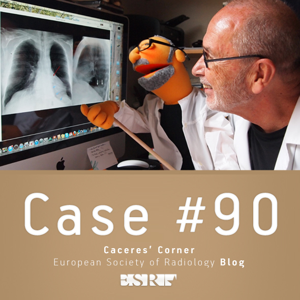
Caceres’ Corner Case 90 (Update: Solution)

ESR_2012_Blog-CaceresCorner-590-CASE90

Dear Friends,

This week we are presenting pre-op radiographs of a 71-year-old man with torn ligaments of the right shoulder. As you can see, an unsuspected abnormality was found. What would be your diagnosis?

Check the images below, leave your thoughts in the comments section and come back on Friday for the answer.

1. Hydatid cyst
2. Carcinoma of the lung
3. Fibrous tumour of pleura
4. None of the above

Read more…

07
Apr 2014
POSTED BY
POSTED IN
DISCUSSION 29 Comments

ESR initiatives to strengthen the visibility and role of imaging in personalised medicine

ECR2014_ECRToday_Blog_Final

An interview with Prof. Hans-Ulrich Kauczor, Heidelberg/DE, ESR Research Committee Chair

ECR Today: What are the Research Committee’s objectives?
Hans-Ulrich Kauczor: Our main tasks are to periodically survey the needs of the research community, as well as develop recommendations for radiologists to engage with different fields of research and innovation. We also work to assess the current status and develop recommendations on how to improve education in research and to provide strategic recommendations for the research field to the executive council. Last but not least, we aim to leverage cooperation between research disciplines and foster networking and liaising with scientific biomedical European societies.

Hans-Ulrich Kauczor is professor and chairman of radiology at the University of Heidelberg and director of diagnostic and interventional radiology at University Hospital Heidelberg in Germany. He chairs the ESR’s Research Committee.

Hans-Ulrich Kauczor is professor and chairman of radiology at the University of Heidelberg and director of diagnostic and interventional radiology at University Hospital Heidelberg in Germany. He chairs the ESR’s Research Committee.

ECRT: Why is it important to have a dedicated ESR subcommittee for imaging biomarkers?
HUK: The Subcommittee on Imaging Biomarkers was established to address the issues concerning the future development of image-derived quantitative biomarkers, its assessment, validation and standardisation.

The development of imaging biomarkers has become an integral part of modern medicine with a huge potential to advance the development of personalised medicine. Different types of imaging biomarkers (anatomical, functional, and molecular) are used for the detection and treatment of major diseases including cancer, cardiovascular diseases, neurological and psychiatric diseases, musculoskeletal disorders, metabolic diseases, as well as inflammatory and autoimmunity based diseases. In contrast to other biomarkers, imaging biomarkers have the advantage of remaining non-invasive. They are also spatially and temporally resolved, non-destructive and repeatable over a long period, and have the potential for broad application. But before imaging biomarkers can be widely adopted, measures for standardisation and quality assurance must be implemented.

Read more…

Dr. Pepe’s Diploma Casebook: Case 54 – SOLVED!

Dr_Pepe_Casebook_54

Dear Friends,

Welcome to this week’s case. Radiographs belong to a 43-year-old male with chest pain. Leave me your thoughts and diagnosis in the comments and come back on Friday for the answer.

Diagnosis:

1. Hypertrophic cardiomyopathy
2. Mitral disease
3. Non-cyanotic congenital heart disease.
4. None of the above

Read more…

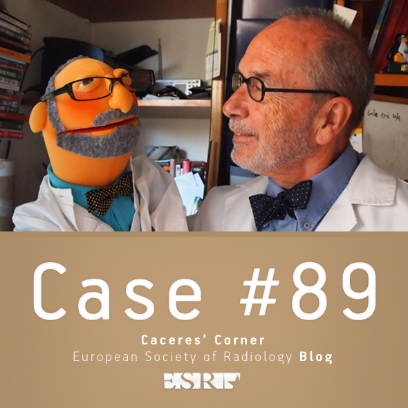
Caceres’ Corner Case 89 (Update: Solution)

ESR_2012_Blog-CaceresCorner-590-CASE89

Dear Friends,

Muppet is concerned about the few responses in the last case. He wants to show an easy case to stimulate participation. Showing pre-op radiographs of a 41-year-old woman with rectal prolapse. The opacity in the right lung was not seen in the CT. What do you think?

Examine the images below, leave me your thoughts and diagnosis in the comments section, and come back on Friday for the answer.

Read more…

24
Mar 2014
POSTED BY
POSTED IN
DISCUSSION 18 Comments

Dr. Pepe’s Diploma Casebook: Case 53 – SOLVED!

Diploma_casebook_case42

Dear Friends,

Today I am presenting radiographs of a 36-year-old woman with shortness of breath increasing over the last three months. Examine the image below and leave me your thoughts and diagnosis in the comments. Come back on Friday for the answer.

Diagnosis:

1. Metastatic disease
2. Sarcoid
3. Interstitial pneumonia
4. None of the above

Read more…

17
Mar 2014
POSTED BY
DISCUSSION 26 Comments

Rising Stars exposes students to “the charms of radiology,” says former ECR President

ECR2014_ECRToday_Blog_Final

ECR Today spoke with Professor Małgorzata Szczerbo-Trojanowska, from Lublin, Poland, patron of the Rising Stars programme.

ECR Today: The Rising Stars programme is under your patronage this year. What motivated you to take on this role?

Malgorzata Szczerbo-Trojanowska: Medical students and residents will create the shape of radiology in the future. Therefore, those of us who care for our specialty should make an effort to encourage the best, brightest and most enthusiastic students to choose diagnostic imaging as their professional career.

I am very pleased to be involved in the preparation of the Rising Stars programme, which aims to create an interesting educational agenda for students and to encourage them to actively participate in the congress sessions specifically designed for them.
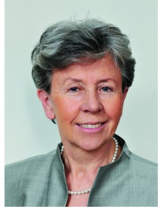
Professor Małgorzata Szczerbo-Trojanowska is Head of the Department of Interventional Radiology and Neuroradiology at the Medical University in Lublin, Poland. She served as ECR Congress President in 2010.

Professor Małgorzata Szczerbo-Trojanowska is Head of the
Department of Interventional Radiology and Neuroradiology at the
Medical University in Lublin, Poland. She served as ECR Congress
President in 2010.

I think this programme is a very important initiative of the European Society of Radiology and the ECR. Having been an academic teacher for many years, I am aware of the great significance of undergraduate education. It has always been my aim as chairperson of the radiology department at my university to offer attractive forms of radiology teaching and to expose students to the charms of radiology from the early years of their education. This is the way to raise their interest in this specialty and get them involved in radiology research. It is in the best interest of the future of radiology. So when I was offered to take patronage of the Rising Stars programme, I didn’t hesitate for even a second.

ECRT: Why should a medical student or radiology trainee take part in this programme?

MST: The European Congress of Radiology, one of the world’s leading radiological meeting, offers medical students and residents a unique opportunity to see the greatest scientists, practitioners, lecturers and teachers present some fascinating achievements of modern radiology. There are also many chances to learn the state of the art in a wide variety of imaging methods for a plethora of diseases, and to find answers to complex problems of contemporary medicine provided by world-renowned experts. Participation in the ECR shows the importance of radiology in patient care, its great impact on other areas of medicine, and its relevance in the progress of medicine.

By taking part in the Rising Stars programme, medical students, radiology trainees and radiography trainees have an opportunity to give a presentation on the results of their own research work and opinions, or ideas, related to medical studies. Visiting the ECR technical exhibition is always an exciting and stimulating experience, providing a unique opportunity to become acquainted with cutting-edge technologies and new solutions.

Read more…