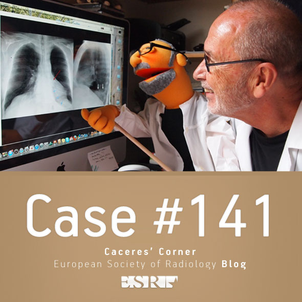
Caceres’ Corner Case 145 (Update: Solution)

esr_2016_blog-cacerescorner-145

Dear Friends,

Today we are presenting a recent case. The PA radiograph was made for routine check up of a 49-year-old male.

Check the image below, leave your thoughts in the comments and come back on Friday for the answer.

Diagnosis:

1. McLeod syndrome
2. Changes post-TB
3. Congenital lung hypoplasia
4. None of the above

Read more…

17
Oct 2016
POSTED BY
POSTED IN
DISCUSSION 12 Comments

Interview: Dr. Sophie Dellas, head of breast imaging and diagnostics at the University Hospital Basel, Switzerland

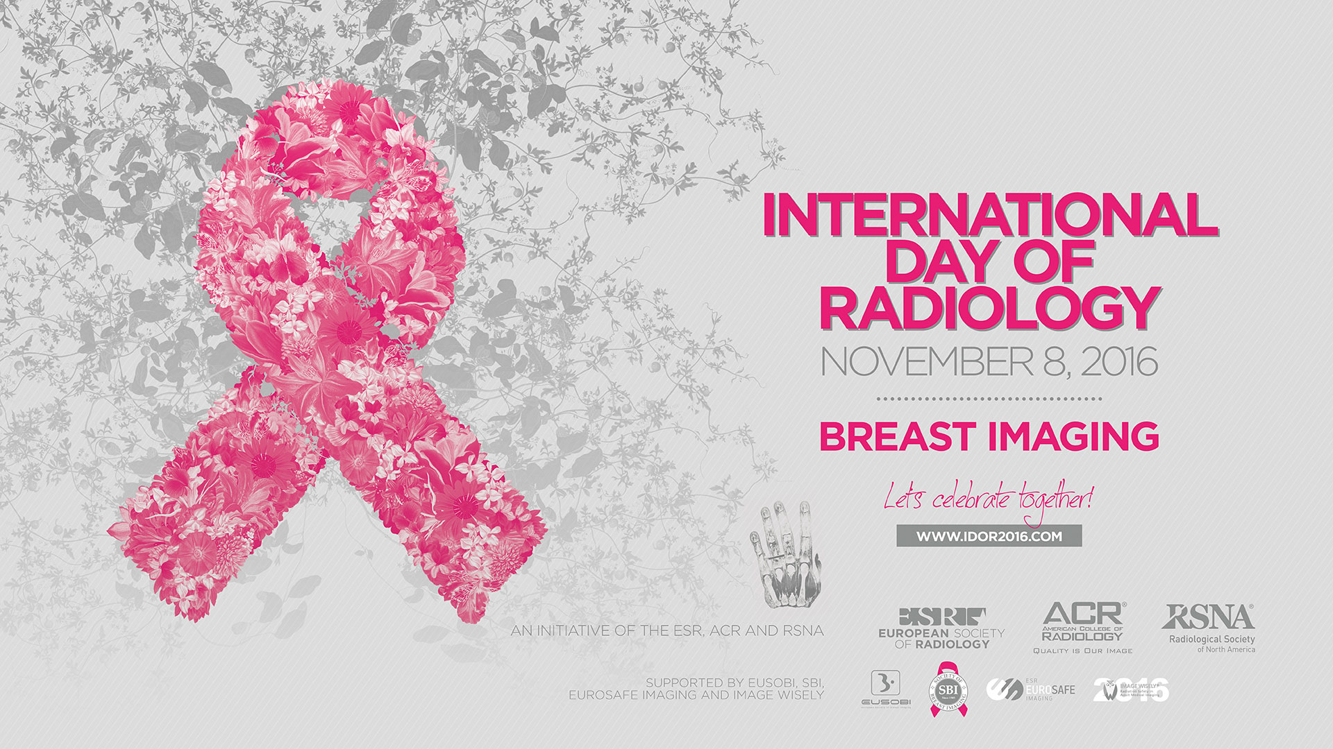
idor_2016_facebook_1920x1080px_72dpi

This year, the main theme of the International Day of Radiology is breast imaging. To get some insight into the field, we spoke to Dr. Sophie Dellas, assistant professor of radiology and division head of breast imaging and diagnostics at the University Hospital Basel, Switzerland, and a core team member of the certified breast centre at the same institution.

European Society of Radiology: Breast imaging is widely known for its role in the detection of breast cancer. Could you please briefly outline the advantages and disadvantages of the various modalities used in this regard?
Sophie Dellas: Mammography is the imaging modality of choice for breast cancer screening, but also for diagnosis, evaluation, and follow-up of people who have had breast cancer. Long-term results of randomised controlled trials of mammography screening on average show a decrease in breast cancer mortality of 22% in women aged 50 to 74 years. The main problem of mammography is that it is not a perfect method. Mammography generates 2D images based on the density of tissue for penetrating x-rays. The compression of the breast that is required during a mammogram can be uncomfortable. The compression is necessary to reduce overlapping of the breast tissue. A breast cancer can be hidden in the overlapping tissue and not visible on the mammogram. This is called a false negative mammogram. Mammography is associated with a false negative rate in the order of 10% to 20%. On the other hand, mammography can identify an abnormality that looks like a cancer, but turns out to be normal. This is called a false positive mammogram. Besides worrying about being diagnosed with breast cancer, a false positive means more tests and follow-up examinations. Furthermore, at least some of the cancers found with screening mammography would never otherwise be diagnosed in a patient’s lifetime. The magnitude of such overdiagnosis is a topic of much debate. It is likely to represent up to 10% of breast cancers found on screening mammography and results in potentially unnecessary treatments.

dellas-sophie_portrait_idor2016

Dr. Sophie Dellas, assistant professor of radiology and division head of breast imaging and diagnostics at the University Hospital Basel, Switzerland.

Breast ultrasound is complementary to both mammography and magnetic resonance imaging (MRI) of the breast. It does not use radiation. It is therefore the initial diagnostic method of choice if breast imaging is required below the age of 40. It allows the confident characterisation of not only benign cysts but also benign and malignant solid masses and the characterisation of palpable abnormalities. The high spatial and contrast resolution of modern breast ultrasound equipment allows the detection of subtle lesions at the size of terminal duct lobular units such as DCIS and small invasive cancers. In women with dense breasts and a negative mammogram, ultrasound therefore is increasingly used as a supplemental screening tool. The major disadvantage of ultrasound as a screening tool is the high risk of false positive findings resulting in unnecessary biopsies. The rate of false positives is much higher with screening ultrasound than with mammography or screening MRI.

Unlike mammography, MRI of the breast does not use radiation. It is safe even though it does require an intravenous injection of a contrast medium. It has a sensitivity exceeding 90% for detecting breast cancer and is superior to mammography and ultrasound. Annual MRI screening is recommended for women with a high lifetime risk of getting breast cancer. Although breast MR imaging is extremely sensitive, its specificity is limited, leading to additional workups and benign biopsies. Good quality breast MR imaging is expensive, time-consuming, and not universally available. Patients with pacemakers, certain aneurysm clips or other metallic hardware, an allergy to contrast agents, or severe claustrophobia are unable to undergo MR imaging.

Read more…

Dr. Pepe’s Diploma Casebook: Case 96 – A painless approach to interpretation (Chapter 3) – SOLVED!

diploma_casebook_case96

Dear Friends,

I hope you remember the three questions to ask when facing a chest radiograph:

a) Is there any visible abnormality? (Chapter 1)
b) Is it intra or extrapulmonary? (Chapter 2)
c) What does it look like?

To discuss the third question, I’m showing chest radiographs of a 36-year-old woman with chest pain.

What does the lesion look like?

1. Pericardial fat pad
2. Thymic tumour
3. Pericardial cyst
4. Any of the above

Check the two images below, leave your thoughts in the comments section and come back on Friday for the solution.

Read more…

Caceres’ Corner Case 144 (Update: Solution)

esr_2016_blog-cacerescorner-144

Dear Friends,

Today we are showing another pre-op case, including a PA chest radiograph of a 62-year-old man with lumbar hernia. Check the image below, give us your thoughts in the comments, and come back on Friday for the answer.

Diagnosis:

1. TB
2. Carcinoma
3. Pulmonary hypertension
4. None of the above

Read more…

03
Oct 2016
POSTED BY
POSTED IN
DISCUSSION 16 Comments

Dr. Pepe’s Diploma Casebook: Case 95 – A painless approach to interpretation (Chapter 2) – SOLVED!

diploma_casebook_case95

Dear Friends,

As you may remember from Diploma Case 94, when I’m facing a chest radiograph I start by asking three questions:

a) Is there any visible abnormality?
b) It is intra- or extrapulmonary?
c) What does it look like?

Today we will discuss the second question, showing chest radiographs in two different patients. Is the abnormality intra- or extrapulmonary? Check the images below, leave your thoughts in the comments and come back for the answer and discussion on Friday.

Read more…

Caceres’ Corner Case 143 (Update: Solution)

esr_2016_blog-cacerescorner-143

Dear Friends,

Today we are presenting pre-op radiographs for inguinal hernia in a 37-year-old-man. Check the images below, leave your thoughts in the comments section, and come back on Friday for the answer.

Diagnosis:

1. Pulmonary arterial hypertension
2. Thymoma
3. Pericardial cyst
4. None of the above

Read more…

19
Sep 2016
POSTED BY
POSTED IN
DISCUSSION 11 Comments

Dr. Pepe’s Diploma Casebook: Case 94 – A painless approach to interpretation (Chapter 1) – SOLVED!

Diploma_casebook_case94

Dear Friends,

This year I intend to discuss the basic principles of interpreting chest radiographs, under the heading, “The Beauty of Basic Knowledge”.

I plan to structure the discussion into three main parts, which will take us through the whole academic year:

Part 1. A painless approach to interpretation
Part 2. To err is human: how to avoid slipping up
Part 3. The wisdom of Dr. Pepe

This week’s case is the first chapter of my ‘painless approach to interpretation’. Interpreting chest radiographs is not difficult if we follow Confucius’ saying: “ A journey of a thousand miles begins with a single step”. As a clinical radiologist, my first step is to ask myself three questions:

a) Is there any visible abnormality?
b) It is intra or extrapulmonary?
c) What does it look like?

Today I’ll discuss the first question. Below you can see the chest radiographs of three different patients. Do you see any visible abnormality in any of them? Let me know in the comments section and come back on Friday for my answer.

Read more…

Caceres’ Corner Case 142 (Update: Solution)

ESR_2016_Blog-CaceresCorner-142

Dear Friends,

Welcome back! To start the academic year I would like to show a case seen by Dr. Pepe while I was vacationing in Minorca.

Radiographs belong to a 35-year-old woman with pain in the mandible for the last two years. CT of the mandible before biopsy and pre-op PA chest radiograph are shown. After seeing both, Dr Pepe suggested a diagnosis. What do you think?

Check the images below, leave your thoughts in the comments section, and come back on Friday for the answer.

Read more…

05
Sep 2016
POSTED BY
POSTED IN
DISCUSSION 23 Comments

Dr. Pepe’s Diploma Casebook: Case 93

Diploma_casebook_case93

Dear Friends

This is the last case of the first semester. It has been a hard year and I need some quiet time. Will see you again on Monday, September 5 with new cases. Relax and enjoy your vacation!

This week’s case follows the pattern of a ‘Meet the Examiner’ presentation, with questions and answers similar to a real examination. Take your time before scrolling down for the answer. And no peeking!

The images were obtained during routine CT screening in a 72-year-old man, heavy smoker.

What would be your diagnosis?

1. Carcinoma in LUL and cyst in LLL
2. Carcinoma in LUL and cystic carcinoma in LLL
3. Tuberculosis
4. None of the above

Read more…

13
Jun 2016
POSTED BY
DISCUSSION 8 Comments

Caceres’ Corner Case 141 (Update: Solution)

ESR_2016_Blog-CaceresCorner-141

Dear Friends,

Showing today pre-op radiographs for a hip prosthesis in a 62-year-old man.
What do you see?

Check the images below, leave your thoughts in the comments section, and come back on Friday for the answer.

Read more…

06
Jun 2016
POSTED BY
POSTED IN
DISCUSSION 24 Comments