Caceres’ Corner Case 182 (Update: Solution!)

Dear Friends,

Today I am showing chest radiographs of a 52-year-old man with liver cirrhosis and moderate dyspnoea. What do you see?

Check out the images below, leave your thoughts in the comments section, and come back on Friday for the answer.

Read more…

16
Apr 2018
POSTED BY
POSTED IN
DISCUSSION 7 Comments

Caceres’ Corner Case 181 (Update: Solution!)

Dear Friends,

Today I am presenting a recent case, seen last January. Below is a preoperative PA chest radiograph for bariatric surgery in a 44-year-old woman. Check the image and leave me your thoughts in the comments section. I will show more images on Wednesday, followed by the final answer on Friday.

What do you see?

Read more…

02
Apr 2018
POSTED BY
POSTED IN
DISCUSSION 17 Comments

ECR 2018 Cases of the Day Winners

The winners of the ECR 2018 Cases of the Day Quiz are as follows:

Mehmet Serindere; Hatay/TR
Sungeun Park; Seoul/KR
Seungchul Han; Seoul/KR
Sewoo Kim; Seoul/KR
S. A. Sohaib; Sutton/UK
Rafal Darecki; Koscierzyna/PL

To view the cases please click here.

Congratulations to all winners!

26
Mar 2018
POSTED BY
POSTED IN ,
DISCUSSION 0 Comments

The best submissions for the “Normal variant or disease” interlude at ECR 2018

Dear Friends,

Over the last couple of years, one of the last sessions at the ECR has always covered 20 interesting cases from various subspecialties, which the audience is asked to solve in an interactive way to broaden and update their knowledge.

In between, the very best submissions from the global radiological community have been presented in an interlude lecture. The best submission has always been awarded with a prize and a certificate.

Due to time limits, not all submitted cases can actually be shown onsite, but the session’s rising popularity has resulted in increasing numbers of submissions of excellent quality and didactic value. This is why we would like to give our submitters the opportunity to reach a broader audience by posting the best cases here on the ESR Blog.

This year’s topic was Normal variant or disease to avoid misdiagnosis, overdiagnosis, unnecessary and costly work-up, as well as patient’s concerns.

When we think back of our time in medical school and remember our studies of embryology, we have probably all thought at some stage, how astonishing it is that a viable fetus develops and a healthy newborn is delivered. Embryological development is so complex that an awful lot can go wrong at any stage. Fortunately, real malformations are rare. In our professional life as radiologists, though, we are not infrequently dealing with normal variants, when development is arrested at a certain stage or turns the wrong way round. Even first year residents are familiar with right sided aortic arch, azygos lobe and accessory ossification centres. Over time, one encounters a whole array of normal variants and learns to interprete them as such. Of course, one can only identify, what one has learnt to see and there is a large spectrum of slight developmental abnormalities, which should not be misdiagnosed as abnormality in need of further imaging, invasive diagnostic procedures or treatment. On the other hand, some developmental disorders have clinical significance and you could be the first to advise the patient.

Below, we are pleased to present the best submissions for the “Normal variant or disease” interlude at ECR 2018:

Caceres’ Corner Case 180 (Update: Solution!)

Dear Friends,

This week’s radiographs belong to a 68-year-old man with pulmonary fibrosis and dyspnoea. Check the images below and leave your thoughts in the comments section. New images will be added on Wednesday, followed by the answer on Friday.

What do you see?
Read more…

19
Mar 2018
POSTED BY
POSTED IN
DISCUSSION 9 Comments

Caceres’ Corner Case 179 (Update: Solution!)

Dear Friends,

I want to present today a case that I saw two months ago. The radiographs belong to an asymptomatic 55-year-old man. Will show more images on Wednesday.

Check the images below, leave your thoughts in the comments section, and come back for the answer on Friday.
Read more…

05
Mar 2018
POSTED BY
POSTED IN
DISCUSSION 15 Comments

Incoming ESR President Lorenzo Derchi looks forward to celebrating an ECR milestone in 2019


By Julia Patuzzi

As has been our tradition for a few years now on the final day of our congress, we already look ahead to next year’s ECR. We therefore spoke with Prof. Lorenzo E. Derchi from Genoa, Italy, who, as incoming ESR President, is in charge of ECR 2019. He told us about his plans and ideas for the next European Congress of Radiology.

ECR Today: Professor Derchi, next year’s European Congress of Radiology will be the 25th to be held in Vienna. Are there any specific celebrations planned that you can already share with us?

Prof. Lorenzo E. Derchi

As incoming ESR president, Prof. Lorenzo E. Derchi, Head of the Department of Radiology at the University of Genoa, will preside over ECR 2019.

Lorenzo E. Derchi: We are still thinking about them. An anniversary like this is something more than an opportunity for a big celebration. It is the right moment to re-think our history and what we have reached over the years. To always have the congress in the same city since 1991, at first every other year and then, after 1999, annually, has been a radical change. The new formula proved successful and ECR has become the European meeting to come to in order to learn the latest clinical and technical advances in our discipline as well as to present the results of the previous year’s research.

In my experience, however, it has become much more. From 1991 up until today all congress chairpersons have been part of the same team in a relay race, each handing off the baton to the next one. This continuity can be felt in the special atmosphere at each meeting, and has allowed the congress to become the annual appointment to meet old friends and make new ones; to discuss any radiological topic and then to start working together on it; and finally a place to meet periodically to share problems, advancements and results of long-term projects.

ECRT: Were you here for the first ECR in Vienna in 1991? If not, when did you attend your first ECR? What do you recall from that visit?

Read more…

Trojanowska to deliver honorary lecture on neglected form of cancer

Polish radiologist Dr. Agnieszka Trojanowska will shed light on human papilloma virus (HPV)-induced squamous cell cancer of the head and neck, a common yet long neglected type of cancer. In an interview with ECR Today, she also explained the origins of her passion for head and neck imaging, and how radiologists will increasingly benefit from computer science.

Dr. Agnieszka Trojanowska, assistant professor in the department of radiology and nuclear medicine at Lublin University Hospital, is an internationally recognised specialist in head and neck imaging. Her special interest for oncology and her natural curiosity led her to agree to deliver the Josef Lissner Honorary Lecture on HPV-induced squamous cell cancer of the head and neck (HNSCC) at ECR 2018.

Agnieszka Trojanowska from Lublin, Poland, will speak on HPV-induced squamous cell cancer of the head and neck in her Honorary Lecture today.

“HNSCC behaves differently than more well-known forms of SCC, and it is becoming more and more prevalent. It affects young, well-educated people with high socioeconomic status, and is a quickly rising sexually transmitted entity with peculiar clinical and molecular characteristics. In particular the rise of HPV-induced cancer has been observed in the United States since the beginning of 21st century,” she said.

It was important for her to talk about this long overlooked topic, for which much remains to be done. “It has been under-estimated for many years in many countries. Nowadays, we can speak about an HPV epidemic, leading to a significant rise of oropharyngeal cancer incidences worldwide. These cancers are quite unique and, in my opinion, knowledge of their physiology and treatment options is essential,” she said.

New research shows that, compared with environmental-related head and neck squamous cell carcinomas, patients with HPV-related malignancies display a better response to treatment and a lower risk of death and progression, Dr. Trojanowska pointed out. It is also noteworthy that, according to the 2017 TNM classification, HPV+ cancers are evaluated with a separate grading scale, she added.

Read more…

The past, the present and the future – a foray through Portuguese radiology


by Katharina Miedzinska

Saturday’s ‘ESR meets Portugal’ session is a successful combination of science, culture and entertainment, offering the opportunity to discover how Portugal has contributed to the practice of radiology over the years and foresee the future of Portuguese radiology.

In an interview with ECR Today, Dr. Filipe Caseiro Alves, professor at the University Clinic of Radiology, Coimbra University Hospitals, Portugal, outlined some central themes and highlights of Saturday’s highly anticipated session.

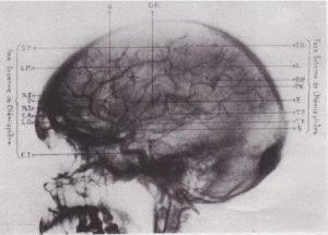
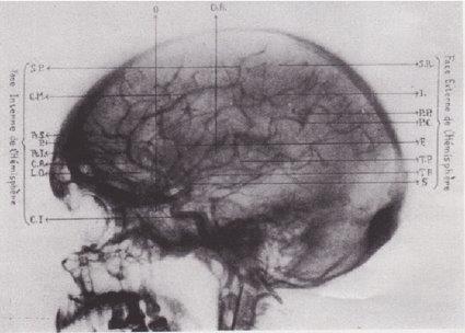
One of the first reports and image of cerebral angiography. From, Moniz E, de Carvalho L, Lima A. La radioartériographie et la topo-graphie cranioencephalique. J Radiol Electrol Med Nucl 1928;12:72. (Provided by Prof. Filipe Caseiro Alves)

At the beginning, Caseiro Alves plans to go on a short time travel through the history of Portuguese radiology. “Portugal’s greatest contributions to radiology are closely interwoven with the Portuguese School of Angiography, which began with the work of Egas Moniz, who was the first to perform a cerebral arteriography in 1927,” he explained.

Since then, Portugal has yielded many outstanding pioneers in the field of vascular radiology, among them Reynaldo dos Santos, a professor of surgery in Lisbon, who, among others, is known for the invention of aortography in 1929. Others include Lopo de Carvalho, who successfully introduced pulmonary angiography in 1931, Álvaro Rodrigues, Sousa Pereira and Roberto de Carvalho, who are known for their innovative work on lymphography (1933), Reynaldo’s son, João Cid dos Santos, who successfully introduced direct phlebography of the limbs in 1938, and Ayres de Sousa, whose name will always be closely associated with microangiography. “All these pioneer works were at the forefront of one of the most important achievements in healthcare and proved instrumental in shaping today’s medical practice,” Caseiro Alves said. Read more…

ECR continues to think outside the box and adds the Cube

For young physicians, interventional radiology (IR) may seem as enigmatic as a Rubik’s cube. But the cube can be solved with the right algorithm, according to Dr. Maximilian de Bucourt, head of angiography at Charité Campus Benjamin Franklin in Berlin.

“What we do in IR is exactly like a Rubik’s cube: you follow the algorithm and solve the problem. In IR, if you do the procedure steps over and over again and use the rules, most of the time you can get the solution for the patient,” he said.

Dr. Maximilian de Bucourt is head of angiography at Charité Campus Benjamin Franklin in Berlin, and one of the specialists behind the Cube.

Together with Prof. Christian Loewe from Vienna, de Bucourt imagined ‘the Cube’, a workshop aimed at introducing young physicians to the tools and techniques used in IR. With its focus on hands-on activities, including simulated procedures and interactive demonstrations, the Cube is fulfilling its goal of acquainting residents with this unique sub-specialty.

“The Cube is for young people who are thinking of becoming IR specialists, but find it too long before they can deploy a stent or manoeuver a catheter inside an artery. After medical school, basic radiology training and dedicated IR training with a teacher willing to let them perform major interventions, they still need to learn how to manage complications. The Cube is all about expediting this process, by enabling students and residents to get their hands on the products earlier,” he explained. Read more…